During poster session 1 of the European Association of Urology (EAU) Congress, Barcelona, Spain, the abstract ‘High field single subject brain mapping of pelvic floor motor control. A 7-Tesla fMRI study’ was presented. The aim of the study was to further define the brain areas involved in pelvic floor motor control and to indicate whether single-subject mapping of urogenital control is possible using 7-Tesla functional MRI (fMRI). Arguments for the aim of this study are the poorly understood pathophysiology of many functional pelvic floor disorders and its high prevalence. PET, 1.5, and 3-Tesla fMRI studies have already demonstrated brain areas to be involved in pelvic floor motor control, but these studies produced inconsistent findings and only group results have been analysed to date. Firstly, the authors hope to further clarify the brain areas involved in this pelvic floor motor control in healthy volunteers, and, secondly, demonstrate that high-field imaging is a suitable technique to detect urogenital control in these individuals.
Seventeen healthy males had undergone a 7-Tesla fMRI (Philips Achieva, Philips, Guildford, UK) performing two tasks: repetitive pelvic floor muscle contractions and, as a control, horizontal tongue movements. Volunteers practised the tasks in the mock scanner. The data from four subjects had to be rejected due to motion artefacts.
During pelvic floor muscle contractions, active clusters were found on the primary motor cortex (M1), supplementary motor area, anterior insula, putamen, thalamus, and cerebellum. Figure 1 shows the results of our group, specifically the individual analyses of both cortex and cerebellar results.
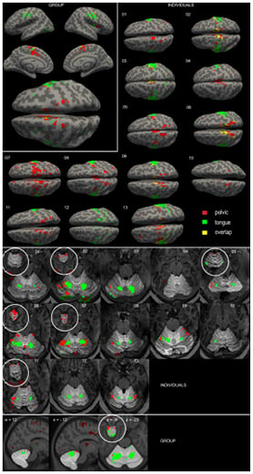
Figure 1: Active clusters found during groups analysis and in individuals of the cortex and the cerebellum.
In all the subjects, activation of the primary motor cortex was visualised, and when compared to the homunculus the activation is located in the hip region. In the group analyses the cluster is split in two, whereas in single subjects it appears to be one cluster. In the anterior lobe of the cerebellum, activation during both tasks was objectified in the group analyses. Moreover, these results were also visible in most of the single subjects.
This study demonstrated which brain areas are involved in pelvic floor motor control, and furthermore, showed that the 7-Tesla fMRI is a suitable technique for brain mapping urogenital control in single subjects. Single subject results were comparable and complementary to group results. The discussion following the presentation of this subject mainly focussed on the clinical applicability of this study. This study indicates that the 7-Tesla fMRI might be suitable for demonstrating differences between healthy subjects and patients with pelvic floor disorders and be able to provide information on the pathophysiology of this highly prevalent disease. Other studies using fMRI have shown disease-related changes in activity patterns in patients with urge urinary incontinence.1 Future perspectives include the clinical potential to use fMRI as a diagnostic tool or to score treatment effects.