Nicholas Fuggle | Associate Professor in Rheumatology, University of Southampton, UK; Alan Turing Institute Clinical AI Interest Group co-organiser
Citation: EMJ Rheumatol. 2025;12[1]:63-65. https://doi.org/10.33590/emjrheumatol/FAJF2950
![]()
EMJ is delighted to introduce Nicholas Fuggle, Associate Professor in Rheumatology at the University of Southampton, UK, and co-organiser of the Alan Turing Institute Clinical AI Interest Group. In this interview, he discusses the evolution of clinical AI, its impact on rheumatology and musculoskeletal research, and how machine learning is transforming disease prediction and treatment.
Could you share the journey that led you to become a Turing Fellow at the Alan Turing Institute, and what impact has this fellowship had on your research?
Dating back to my PhD, I used machine learning to analyse epigenetic datasets, which sparked my interest in the field. Then, a call went out for Turing Fellows from the University of Southampton, and I was fortunate to be awarded the fellowship, which allowed me to continue building relationships with colleagues at the Turing Institute, as well as with other researchers across a diverse range of specialties. This ultimately led to me co-founding the Clinical AI Interest Group in May 2022.
Now, with over 1,300 members, the group serves three key purposes: fostering discussions on clinical AI, providing a pool of resources to feed into Turing-related activities, and advancing education. We emphasise that for AI to be effectively integrated into clinical practice, clinicians must understand it well enough to explain its impact to patients. To support this, we run initiatives like a Summer School in Clinical AI to enhance clinicians’ knowledge.
How has the field of clinical AI evolved during your career, and what developments have been most transformative for healthcare professionals?
It’s fascinating to look back at the history of clinical AI. Many don’t realise that the first computer vision paper on osteoporosis dates back to 1996. AI has cycled through periods of excitement (“AI summers”) and setbacks (“AI winters”), but I believe the current AI summer is here to stay for three key reasons.
First, massive datasets like the Picture Archiving and Communications System (PACS) that provide rich training data for AI models. Secondly, advances in hardware, including cloud computing and graphics processing units (GPUs), have made it possible to analyse these datasets in a time-efficient manner. Thirdly, model architectures, with developments like the transformer model behind large language models such as the generative pre-trained transformer (GPT), have reduced the computational burden required to build sophisticated AI tools, and those three reasons suggest that clinical AI is set to remain now.
My own interest in AI began in 2015 after watching a TED Talk on computer vision. At the time, most clinical AI buzz cantered around mammography, aiming to ease the burden of breast cancer screening. Computer vision remains the most advanced area due to the vast amount of imaging data available. However, large language models and ambient consultation recordings are rapidly catching up. While we haven’t yet hit an inflection point, we may soon see language models overtake computer vision in clinical AI applications. It’s an exciting space, and I’d encourage others to get involved.
Predictive modelling is a significant strength of AI. How can it be used to anticipate disease progression or treatment outcomes in rheumatology?
In clinical practice, particularly in screening, we aim to predict disease occurrence or health changes early to modify risk and improve outcomes.
In my specialist area of osteoporosis, we use the Fracture Risk Assessment Tool (FRAX), a predictive tool built on conventional statistical methods rather than AI or machine learning (ML). Its advantage lies in being entirely explainable. However, AI holds great potential in areas like predicting rheumatoid arthritis flares or identifying which patients will respond best to treatments.
I think that AI and ML have a role to play in identifying patterns that we as clinicians aren’t able to see in patient data, including their electronic health records, blood tests, and imaging, taking all of that information together in a multi-modal way, and then leveraging the developments in AI to then predict future outcomes. Supervised machine learning, where models are trained on labelled datasets (e.g., patients who develop flares versus those who don’t), is particularly useful in this context.
What challenges have you encountered when deploying computer vision for imaging in musculoskeletal research?
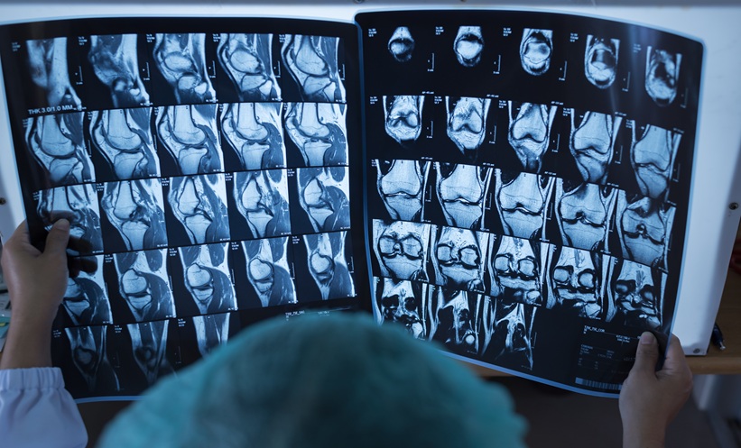
There are several challenges, both from a research and clinical perspective. In rheumatology, imaging helps to identify facets of disease. If we’re looking at a knee with osteoarthritis, for example, we’re assessing for the presence of subchondral sclerosis, bony changes, osteophyte formation, and joint space narrowing. All of these features help guide diagnosis and also allow us to classify the severity of the disease.
A key challenge in computer vision research is the variability in imaging data, dual-energy X-ray absorptiometry scans, for example, can differ across manufacturers, affecting comparability and potentially leading to misinterpretations of bone mineral density. This inconsistency impacts both research and clinical decision-making.
Another issue is variability in expert interpretation. Radiologists may disagree on findings, making it difficult to establish a definitive “gold standard.” If expert assessments are inconsistent, training AI models to replicate them becomes challenging. These are among the biggest hurdles in deploying AI for musculoskeletal imaging.
What do you think are the main barriers to integrating AI tools into everyday clinical workflows, and how can healthcare systems address them?
The barriers are substantial and vary depending on the healthcare system. One key issue is ensuring AI tools are both effective and safe for deployment. Another is determining where they fit within existing workflows. For example, AI can opportunistically assess bone mineral density from X-rays, but where should this be implemented? In regions without dual-energy X-ray absorptiometry access, such as parts of Africa, or as an automatic feature for all hospital X-rays? While promising, this could increase clinician workload, requiring result interpretation, treatment decisions, and patient consent.
Regulation is another major (though important and entirely necessary) hurdle; AI tools need ongoing quality assessment. Automation bias is also a concern, as clinicians must balance AI insights with their own judgment. AI should enhance decision-making. Addressing these barriers is key to successful integration.
Lastly, how do you envision the role of AI evolving in musculoskeletal research and rheumatology over the next 10 years?
My predictions are that in the next decade, AI will likely play a key role in musculoskeletal research and rheumatology. One major advancement will be the opportunistic identification of vertebral fractures and low bone mineral density, improving osteoporosis screening and fracture risk assessment.
In inflammatory arthritis, AI will increasingly predict flares by integrating electronic health records, omic data, imaging, and clinical outcomes. This multimodal approach could help identify at-risk individuals and tailor treatments more effectively.
In research, machine learning will refine disease classification, identifying distinct endotypes within broad diagnoses like rheumatoid arthritis. This could lead to more precise classifications based on treatment response or flare risk, ultimately benefiting patients.