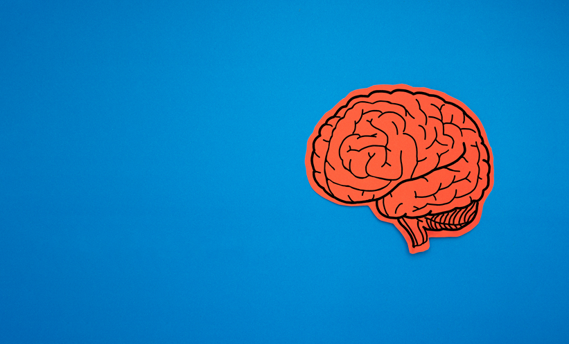
A PIONEERING study has shed new light on the brain structures critical to human verbal memory, offering valuable insights for treating patients with memory impairments, particularly those with temporal lobe epilepsy (TLE). Researchers have identified key cortical and hippocampal regions that play a crucial role in verbal memory encoding, findings that may shape future therapeutic strategies.
The cross-sectional study, involving 84 patients with radiologically and pathologically confirmed hippocampal sclerosis (HS) and unilateral TLE, along with 43 healthy volunteers, examined the relationship between brain structure and verbal memory performance. Using high-resolution MRI scans, researchers extracted morphometric and volumetric measures of the cerebral cortex and hippocampal subfields. Standardised neuropsychological evaluations assessed verbal memory function.
The study’s results highlight a strong association between reduced grey matter volume in specific brain regions and impaired verbal memory. Decreases in the medial and dorsolateral prefrontal cortex, superior and middle temporal gyri, and anterior and posterior cingulate cortex were particularly linked to verbal memory deficits (Pcorr < 0.0001). Additionally, reductions in the left ventrolateral prefrontal cortex and the parietal-temporal-occipital junction were similarly associated with poor verbal learning (Pcorr < 0.0001).
Within the hippocampus, diminished volumes in the left dentate gyrus (P = 0.003), cornu ammonis 4 (P = 0.005), and cornu ammonis 3 (P = 0.03) were found to negatively impact verbal memory scores. These findings were consistent across the entire cohort and within the subgroup of patients with HS, reinforcing the significance of these structures in verbal learning processes.
The research provides compelling evidence of a corticohippocampal network crucial for verbal memory in TLE patients. This understanding could guide future interventions, including neuroprotective strategies to preserve verbal memory following epilepsy surgery. Scientists suggest that preserving parts of the left dentate gyrus, cornu ammonis 4, and cornu ammonis 3 during surgical procedures may help mitigate post-operative verbal memory impairment.
With verbal memory playing a vital role in communication and daily life, these findings represent an important step towards targeted therapies. Future research will explore ways to harness this knowledge for clinical applications, potentially improving the quality of life for epilepsy patients worldwide.
Reference
Fiore G et al. Cortico-hippocampal networks underpin verbal memory encoding in temporal lobe epilepsy. Brain Comms. 2025;7(2):fcaf067.