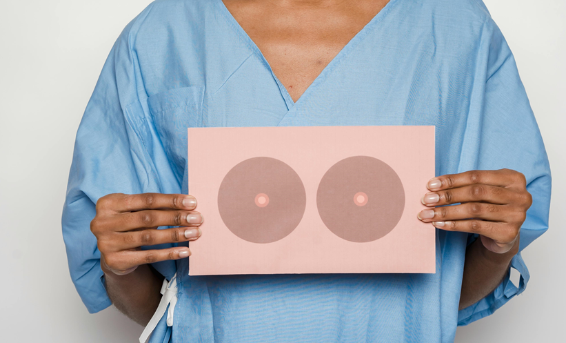
DEEP learning methods have the potential to improve clinical strategies for addressing ultrasound Breast imaging-reporting and data system (BI-RADS) 4A lesions, according to recent research.
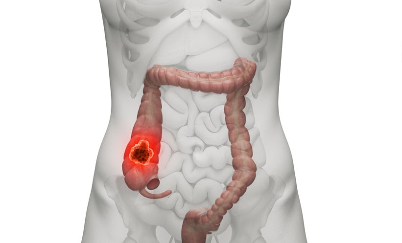
Typically, biopsy is recommended for ultrasound BI-RADS 4A lesions; however, researchers have noted that the malignancy rate of these lesions ranges from 2–10%, and therefore more efficient assessment techniques to distinguish between benign and malignant lesions could improve clinical decision-making. Previous studies have demonstrated the usefulness of deep learning models in breast cancer screening; however, there is a lack of relevant data on the application of deep learning to ultrasound BI-RADS 4A lesions.
The team, led by Mei Yi, Southern Medical University, Guangzhou, China, developed a predictive nomogram using a convolutional neural network, based on clinical and ultrasound features. The study involved 1,616 patients from two hospitals, which were randomly divided into training and internal validation cohorts at a ratio of 7:3, with 100 patients from another hospital making up an external validation cohort.
The nomogram achieved higher area under the curve and sensitivity values than clinical experience alone, whilst maintaining specificity. Results demonstrated that the performance of two radiologists involved in the study was significantly improved when assisted by the nomogram, including areas under the curve of 0.801 and 0.800 with assistance, compared to 0.712 and 0.547 with no assistance, respectively. The nomogram showed the same trend for specificity, improving performance from 70.93–74.42% for one radiologist (p<0.001), and from 59.3–81.4% (p=0.004) for the other radiologist. The deep learning model decreased unnecessary biopsies for both radiologists by 6.7% and 24.0% respectively.
These results highlight the value of deep learning in clinical practice, aiding clinicians’ decision-making, and reducing unnecessary biopsies. “Notably, we found that a hyperechoic halo was significantly related to breast cancer, which may be caused by the infiltration of cancerous tissue into the peripheral tissue,” commented the study authors. “Hence, we suggest that BI-RADS 4A breast lesions with hyperechoic halos should be paid high attention to, especially in older women,” they added.