RESEARCHERS have discovered that the progression of grey matter (GM) atrophy in the brain of patients with mild Parkinson’s disease can be predicted by visualising the structural and functional organisation of the brain using MRI.
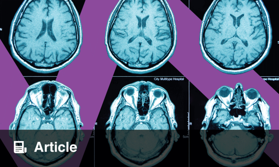
The study, led by Silvia Basaia, Division of Neuroscience, Milan, Italy, involved 86 patients with mild Parkinson’s disease and 60 healthy controls. Three-dimensional T1‑weighted brain MRI scans were used to generate a detailed map of the brain’s neural connections, a brain connectome. From this, they measured the atrophy of neural connections by observing changes in GM at baseline and every year for three years following.
A disease exposure index was calculated and, when correlated with brain atrophy over time, revealed a significant relationship between disease presence at 1 and 2 years and the subsequent atrophy at 2 and 3 years. Models using these indexes, alongside demographic and clinical variables, accurately predicted atrophy in several brain regions suggesting that the connectome could be used to predict GM atrophy.
Previous research into Parkinson’s disease has shown that GM atrophy in similar patient groups is associated with the progression of motor, nonmotor, and cognitive symptoms. Therefore, these results showing successful atrophy prediction suggest that MRI-based brain connectome mapping could serve as a powerful aid of early intervention and personalised treatment for patients with mild Parkinson’s disease.
These findings represent a significant advancement in the field, not only for early clinical identification but also for identifying future biomarkers and further understanding the underlying mechanisms of Parkinson’s disease.
Katie Wright, EMJ
Reference
Basaia S et al. brain connectivity networks constructed using MRI for predicting patterns of atrophy progression in Parkinson’s disease. Radiology. 2024;311(3):e232454.