INTRODUCTION
Antegrade superficial femoral artery puncture is by far the preferred approach in performing below-the-knee (BTK) percutaneous interventions. However, locating the proper arterial site is sometimes difficult, either because of excessive fat deposition in the groin or because of a hardly perceptible pulse due to severe obstructive lesions of the iliac or common femoral vessels. As a result, direct puncture can be frustrating, time-consuming, and often a cause of unjustified procedure prolongation and patient discomfort.
Angiographic visualisation of the target vessel using a different arterial access (contra-lateral leg, upper arm) is a frequently used option. However, this approach consistently prolongs procedure time, along with radiologic exposure; this implies an adjunctive arterial puncture, therefore increasing the likelihood of unnecessary complications and increasing contrast utilisation. As an alternative, the vessel can be visualised by an echo scan, which requires the presence of a dedicated sonographer in the cath lab, or an echo experienced interventionalist,1 conditions that are not necessarily met in all cath labs.
We developed an easy, innovative technique that proved to be successful in the large majority of challenging cases reported here.
METHODS
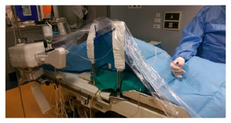
All BTK procedures performed in our centre in the last 5 years have systematically employed an antegrade isocurrent approach, possibly combined with retrograde pedal or posterior tibial puncture in more challenging cases. Furthermore, to optimise timing and contrast utilisation and to minimise embolic complications, diagnostic and interventional procedures are performed using the ACIST–CVi® contrast injection system (ACIST Medical Systems, Inc., Eden Prairie, Minnesota, USA), which provides continuous haemodynamic monitoring and enables real-time pressure reading (Figure 1).

Figure 1: ACIST System connected to a Cook needle through the dye-saline injector/arterial line monitor.
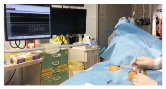
For the arterial puncture, we used a 70 mm 18G Cook needle (B. Braun Melsungen, Melsungen, Germany) directly connected to the arterial line of the CVi device. The needle is advanced through the skin about 1 inch proximally to the inguinal ligament with a 45° angle and a slightly medial orientation pointing at the homolateral big toe. As the needle enters the target vessel, the arterial pressure wave appears on the monitor screen, confirming cannulation (Figure 2). A few millilitres of contrast medium are then injected directly through the needle from the ACIST pump, allowing instant visualisation of the selected vessel. The procedure is then completed by advancing a guidewire according to the standard Seldinger technique.

Figure 2: As soon as the arterial vessel has been cannulated, the arterial line appears all of a sudden on the previously flat curve in the polygraphic monitor.
RESULTS
Out of a total of 276 BTK consecutive procedures performed in the last 5 years, 24 were performed with the standard Seldinger2 approach and 252 with the new smart needle technique. The Seldinger technique was performed essentially by junior interventionalists, before they were trained in the new procedure.
The reasons for choosing the latter were not related to excessive fat deposition, inability to feel the pulse, or inability to enter the artery; all the patients were approached with the same technique due to the high success rate, independent from the anatomical properties.
The success rate of our method has been almost 100%; the only exceptions were vessel occlusions undetected by the preprocedural echo scan or extensive wall calcifications confirmed by angiography performed through the Cook needle; <1% of complications occurred unrelated to the technique but after sheath removal (12 haematomas, 8 pseudoaneurysms, no vessel dissections).
COMMENT
The new smart needle technique has proved to be highly successful and user friendly; the same procedure allows a direct visualisation of the selected artery, thus preventing unplanned cannulation either of the profunda or the common femoral artery. We recommend this easy, cheap, and effective approach in order to simplify the femoral artery puncture both for skilled and for less experienced BTK operators.


