Abstract
Introduction: The presentation of endometriosis as massive haemorrhagic ascites and the presence of concurrent encapsulating sclerosing peritonitis is extremely rare in medical literature. This presentation warrants a strong suspicion for endometriosis especially in a nulliparous, reproductive-age woman.
Case Report: A 22-year-old woman presenting with nonspecific abdominal discomfort, distension, massive ascites, dysmenorrhea, anaemia, and mild weight loss is reported here.
Diagnosis: Based on biopsy results at laparotomy, negative Tuberculosis cultures, a poor response to the anti-tuberculosis therapy, and, finally, an excellent response to combined oral contraceptive pills, endometriosis was confirmed as the diagnosis of exclusion in this case.
Interventions and outcomes: The patient received medical treatment for endometriosis and had an excellent response to treatment.
Methods: The team conducted a thorough literature review on PubMed/MEDLINE, Cochrane, and Science Direct and shortlisted 13 highly relevant articles for the case report. The patient provided informed written consent for the publication of this case report, with no patient-identifying information included in the article. The figures in the case report have not been published elsewhere, so the authors did not require copyright permission.
Conclusion: This is one of the few cases reported in the literature in which endometriosis presented with haemorrhagic ascites and sclerosing peritonitis. Endometriosis should be considered a differential diagnosis in a nulliparous, reproductive-age female who presents with massive recurrent haemorrhagic ascites.
Key Points
1. Overall, 10–15% of all reproductive-age women and 70% of women with chronic pelvic pain have endometriosis. Haemorrhagic ascites as the first presentation of endometriosis is rarely reported.2. This is a case report describing a young woman with recurrent haemorrhagic ascites and an abdominal cocoon found during laparotomy who was diagnosed with endometriosis after 18 months of anti-tuberculosis treatment.
3. The distinct presentation of endometriosis should be considered in the differential diagnosis of women of reproductive age who present with massive recurrent haemorrhagic ascites.
INTRODUCTION
Endometriosis is defined as the presence of endometrial glands and stroma outside the uterine cavity, which commonly presents with subfertility and chronic pelvic pain in reproductive-age women. It affects roughly 10% of the global reproductive-age women’s population. However, this percentage is significantly higher in women with chronic pelvic pain at about 75% and in those with infertility at about 40%.1 Since Brews described the first case of endometriosis presenting as haemorrhagic ascites (HA) in 1954, less than 100 cases have been documented so far.2
The first presentation of endometriosis as recurrent HA is rare in the literature and warrants strong clinical suspicion, especially in young, nulliparous, reproductive-age women of African descent. The criterion for HA is the presence of more than 10,000 red blood cells per microliter. Furthermore, only six cases have been reported in which women presented with both endometriosis-related ascites and encapsulating peritonitis, both of which were present in this case. Encapsulating peritonitis, abdominal cocoon, or frozen ascites is characterised by a fibrin membrane that entraps the bowel loops.3
The authors present the case of a 22-year-old woman with massive recurrent haemorrhagic ascites, first diagnosed on an abdominal ultrasound scan for occasional abdominal discomfort, abdominal distension, mild weight loss, and a thin fibrinous membrane encasing the bowel loop on laparotomy. Endometriosis was subsequently confirmed as the diagnosis of exclusion because of this patient’s excellent response to the combined oestrogen-progesterone oral contraceptive pills. This case provides valuable insights for the consideration of endometriosis as an important differential diagnosis in the evaluation of nulliparous women of childbearing age presenting with massive HA.
CASE PRESENTATION
The case presented here is of a 22-year-old Pakistani woman who presented with abdominal distension, intermittent dull abdominal pain and discomfort, and 8 kg unintentional weight loss in 2 years. Menarche was at age 11 years. She was not sexually active. She had primary dysmenorrhoea but regular menstrual cycles and no history of cyclic pelvic pain. The patient had a past surgical history of an open laparotomy and appendectomy at age 12 years for acute abdominal pain; intra-operatively, a thin fibrinous membrane encasing the bowel loops was found. Based on tuberculous suspicion, histopathology was done then, but no acid-fast bacilli were detected. The surgeon reported no suspicion or findings of endometriosis during this laparotomy.
Apart from past surgical history, there was no significant medical history, and she was not on any regular medication apart from the occasional use of ibuprofen for primary dysmenorrhea. On presentation, physical examination revealed generalised distension but no tenderness. The percussion note was dull, particularly in the lower quadrants.
INVESTIGATIONS
Blood analysis showed microcytic anaemia with a haemoglobin of 11.5 g/dL. An ultrasound scan of the abdomen and pelvis revealed moderate to significant ascites in the pelvis, extending to the right and left paracolic gutters. The uterus and right adnexa were normal; however, there was a haemorrhagic cyst with internal echoes in the left adnexa and the left hydrosalpinx. An ultrasound-guided ascitic tap revealed chocolate-coloured haemorrhagic fluid. Cytological analysis of the ascitic fluid showed numerous red blood cells and some lymphocytes, but there was no evidence of malignant cells. Fluid was exudative with a decreased serum ascitic-albumin gradient. Cultures were negative, and no acid-fast bacilli were detected.
Serum tumour markers demonstrated no significant increase in CA-125 levels. CT of the abdomen and pelvis showed moderate to significant loculated ascites, more marked in the pelvis and extending to the right and left paracolic gutters. The decision for exploratory laparotomy was made to obtain tissue biopsy, as an imaging-guided attempt for biopsy was considered difficult due to the loculated nature of ascites and the presence of peritoneal adhesions because of previous laparotomy.
Findings at laparotomy included multiple adhesions, approximately 4 L of haemorrhagic ascites, a thin fibrinous membrane encasing bowel loops, and a normal-appearing uterus and ovaries. Multiple biopsies of the omentum and ovarian lining were obtained. Histopathology revealed many hemosiderin-laden macrophages and benign-appearing mesothelial cells. There was no evidence of malignant cells. Cultures were negative, and acid-fast bacilli were not detected.
TREATMENT
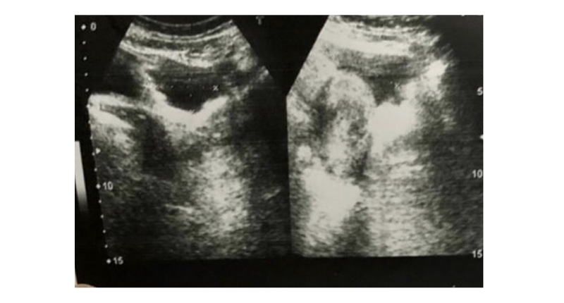
Based on clinical suspicion and the increased burden of disease in the region, peritoneal tuberculosis (TB) was suspected, and empiric treatment of TB was started. The first-line anti-TB therapy (ATT) regimen continued for 1.5 years. However, after 8 months of ATT initiation, imaging, as shown in Figure 1, confirmed the recurrence of ascites, which was drained. After completing the course of ATT, a follow-up MRI of the pelvis revealed a left-sided large hematosalpinx with left paracolic haemorrhagic fluid. There was a small endometrioma in the left ovary and bilateral haemorrhagic follicles. The uterus was unremarkable.
An alternative diagnosis of endometriosis was considered due to the ineffective response to ATT, the lack of objective proof of TB, and an indication of endometriosis on MRI. The patient was started on continuous oestrogen-progesterone oral contraceptive pills.

Figure 1: An ultrasound image of the abdomen and pelvis shows a small amount of fluid in the pelvis after laparotomy and the drainage of ascites.
OUTCOME AND FOLLOW-UP
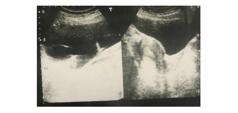
The patient responded well to combination oral contraceptive pills (COCP), and imaging showed decreasing trends in intrabdominal fluid collection. The patient reported no active complaints and was asymptomatic. Continuous COCPs were continued for 1 year, and the patient remained amenorrhoeic. Follow-up imaging revealed no evidence of ascites recurrence; the patient discontinued using COCPs for 3 months and began to have regular menstruation again. However, then she started experiencing discomfort and pain in the left lower quadrant. As shown in Figure 2, the abdominal ultrasound confirmed the re-accumulation of fluid in the left paracolic gutter, which measured around 370 mL. She was then placed back on continuous COCP and has reported no active complaints. She is now under periodic surveillance to look for a recurrence of ascites. The patient is not currently planning for a family; however, she was counselled regarding potential fertility and pregnancy complications in the future. She was also counselled on the possibility of IVF and surgery if needed. Other medical or surgical treatment options can also be considered depending on the patient’s preference.

Figure 2: An ultrasound image of the abdomen and pelvis shows re-accumulation of fluid in the left paracolic gutter after completing the course of anti-tuberculosis therapy for 18 months, prompting a change in treatment to combination oral contraceptive pills, considering endometriosis as the primary diagnosis.
DISCUSSION
Endometriosis is a benign condition in which cells lining the uterus, or endometrium, deposit outside the uterus, causing pain and infertility. Overall, 10–15 % of all reproductive-age women and 70% of those with chronic pelvic pain have endometriosis.4 Endometriosis usually presents with chronic, cyclical pelvic pain, deep dyspareunia, and subfertility; however, recurrent HA as an initial presentation of endometriosis is extremely rare.5
Endometriosis presenting as massive HA is often initially mistaken for peritoneal TB or ovarian or primary peritoneal malignancy as its symptoms of weight loss and decreased appetite often mimic the symptoms in these gynaecological malignancies.6 Additionally, fibroids, Meigs syndrome, benign ovarian tumours, and ovarian hyperstimulation syndrome are among the benign gynaecologic diseases that have been linked to ascites, which makes it challenging to arrive at a final diagnosis.7 In the most recently published systematic review and meta-analysis, the highest prevalence of HA was noted in nulliparous women of African origin. Abdominal distension, weight loss, abdominal pain, and abnormal uterine bleeding were the most common symptoms. In contrast, pelvic mass was the most common physical finding.8 Due to the unusual presentation, it is often dealt with as a diagnostic dilemma, leading to delayed treatment and adding to the patient’s distress. This case emphasises the need for its early recognition and treatment.
The exact cause of endometriosis is not entirely understood; however, various theories exist to explain its pathophysiology. The most postulated theory is that retrograde menstruation of oestrogen-sensitive endometrial cells implanting on peritoneal surfaces elicits an inflammatory response accompanied by angiogenesis, adhesions, fibrosis, scarring, and organ distortion, leading to pain and infertility.7 Furthermore, the precise pathophysiology of endometriosis causing HA and encapsulating peritonitis is unknown; however, according to Bernstein, it may be caused by irritation of the peritoneum by free blood released from ruptured endometrioma, which further enhances fibrosis and inflammation.9 Bloody ascites may be the result of enhanced angiogenesis and friable soft tissue erosions on serosal or peritoneal surfaces, causing micro or frank bleeding. Bloody pleural effusions could be another common finding in patients with HA due to endometriosis, and the most likely cause for this is anatomical abnormalities in the diaphragm.10
The presentation of massive haemothorax is almost always in conjunction with massive ascites. Many of them have diaphragmatic and pleural lesions that need to be surgically repaired.11 The gold standard for diagnosing diaphragmatic endometriosis is video laparoscopy; for thoracic endometriosis, it is video-assisted thoracoscopic surgery.12 Endometriosis risk factors include early age at menarche, shorter menstrual length, and taller height; risk factors associated with a lower risk include smoking, parity, and higher BMI.13 A study by Kaabachi et al.14 showed a statistically significant increase in the expression of IL-37 mRNA in peritoneal fluid in women with endometriosis compared to healthy controls. Moreover, levels of IL-37 mRNA were directly correlated with disease severity.14
Encapsulating sclerosing peritonitis or abdominal cocoon syndrome is defined as a fibro-collagenous membrane surrounding the small bowel or cocoon-like appearance.15 According to a recent systematic review by Magalhães et al.16 on endometriosis-related ascites and encapsulating peritonitis, only six cases of endometriosis-associated encapsulating peritonitis can be found in the literature. The most common primary cause of abdominal cocoon syndrome is idiopathic. However, its secondary causes include endometriosis, peritoneal dialysis, ventriculoperitoneal or peritoneo-venous shunts, liver transplantation, recurrent peritonitis, and familial Mediterranean fever.17 A case report by Yılmaz on a young female patient on haemodialysis revealed diffuse peritoneal thickening and cocoon formation, with recurrent hemoperitoneum corresponding with her menstrual cycles. Endometriosis was further confirmed on biopsy.18 Both endometriosis and encapsulating peritonitis were present in the authors’ case.
CONCLUSION
Despite its rarity, this case highlights the importance of the unique presentation of endometriosis that should be considered in the differential diagnosis of women of reproductive age presenting with massive haemorrhagic ascites.19


