Automatic Method for Jitter Estimation in Electromyographic Signals - European Medical Journal

Automatic Method for Jitter Estimation in Electromyographic Signals

1 Mins
Neurology
Authors:
*Armando Malanda,1 Daniel Stashuk,2 César Valle,1 Javier Rodríguez-Falces,1 Javier Navallas,1 Mamede de Carvalho,3,4 José Castro,3,4 Oscar Garnés-Camarena5
  • 1. Public University of Navarra, Spain
  • 2. University of Waterloo, Canada
  • 3. University of Lisbon, Portugal
  • 4. Santa María Hospital, Lisbon, Portugal
  • 5. Jiménez Díaz Foundation University Hospital, Madrid, Spain
*Correspondence to [email protected]
Disclosure:

This work has been funded by the Department of Health of the Government of Navarra (project GN2022/29) and by the Spanish Ministry of Science and Innovation (project PID2022-136620OB-I00), with payments made to the Public University of Navarra. The authors have declared no other conflict of interest.

Citation:
EMJ Neurol. ;12[1]:58-59. https://doi.org/10.33590/emjneurol/KCDW3797.
Keywords:
Electromyographic, jitter, motor unit potential, neuromuscular junction disease, neuropathies.

Each article is made available under the terms of the Creative Commons Attribution-Non Commercial 4.0 License.

INTRODUCTION

Neurophysiological jitter measurement using concentric needle electrodes is a laborious and difficult task because potentials from several fibres may appear in the recordings due to the large recording areas of these electrodes.1 This work presents the use of an automatic method that estimates jitter from motor unit potential (MUP) trains recorded using concentric needle electrodes.2

METHODS

MUP intervals likely being formed by only one muscle fibre potential were detected from MUP trains. Then jitter measurements were performed between pairs of these intervals. One hundred and thirty-two electromyographic signals were recorded from eight patients with disorders of the neuromuscular junction or different neuropathies, using a Keypoint system and facial-concentric electrodes. These signals were decomposed into several MUP trains using decomposition-based quantitative electromyography software. Jitter measurements of mean absolute consecutive differences (MCD) were then estimated manually, using a home-made graphical interface (Figure 1A and B) and using the automatic method (Figure 1C and D).

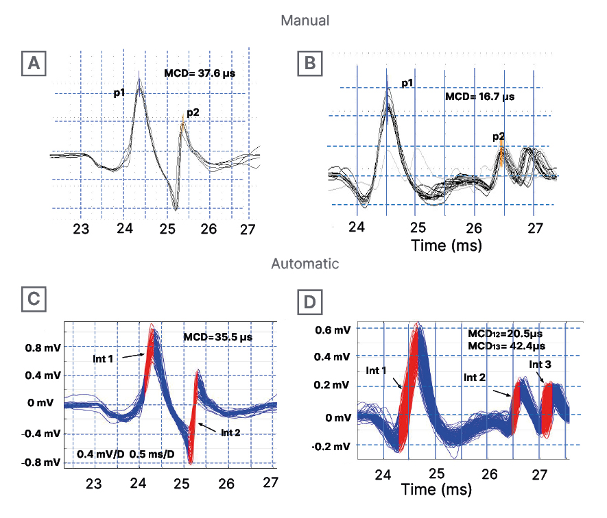
Figure 1: Examples of motor unit potential trains analysed by the manual method, with marked negative peaks (A and B); and by the automatic method, with highlighted valid intervals ​​(C and D).
Obtained MCD values are shown.
MCD: mean absolute consecutive differences; ms: milliseconds.

CONCLUSIONS

The proposed method can be a valuable tool in clinical practice for obtaining reliable automatic jitter measurements and also as a real-time guide for manual jitter measurements. Still, new tests should be carried out to broaden the scope of this study.

References
Sanders DB, et. al. Single fiber electromyography and measuring jitter with concentric needle electrodes.  Muscle Nerve. 2022;66(2):118-30. Malanda A et al. Automatic jitter measurement in needle-detected motor unit potential trains. Comput Biol Med. 2022;149:105973.

Please rate the quality of this content

As you found this content interesting...

Follow us on social media!

We are sorry that this content was not interesting for you!

Let us improve this content!

Tell us how we can improve this content?