There is a well-recognised association between inflammatory dermatomyopathy, dermatomyositis, and underlying visceral malignancy in adults. It is most commonly associated with malignancies arising in the lungs, breasts, and stomach. This is a report of a case found in association with transitional cell carcinoma of the bladder, a site which has only featured in a handful of previous reports.1-8
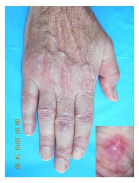
A 68-year-old man, with several weeks history of frequent micturition and haematuria under urological investigation, presented with a history of 3-months’ general illness with muscular pain and weakness, causing him difficulty arising and walking up or down stairs. Physical examination revealed photo-distributed poikiloderma with flagellate erythema of the posterior trunk and nailfold changes (includes ragged cuticles, cuticular hypertrophy, nail-fold telangiectasia) associated with proximal scapular muscular deficit with deltoid amyotrophy (Figure 1). Routine laboratory analysis revealed that creatine phosphokinase levels had reached 1,430 UI/L (normal; 24–195 UI/L), serum lactate dehydrogenase levels were at 544 UI/L, and aldolase A reached 32 UI/L.

Figure 1: Erythematous bands on the back of hands with Gottron’s papules. Upon dermoscopy (lower-right), surface scales and dotted vessels on a homogenous pink background can be seen.
Immunological tests such as the antinuclear antibody test (titer: 320 with speckled nuclear fluorescence), and the anti-TIF1-γ antibody test, were both positive. Electromyography showed increased insertional activity and spontaneous fibrillations with abnormal myopathic low-amplitude, short-duration polyphasic motor unit potential, and complex repetitive discharges in both deltoid muscles. Transurethral cystoscopic examination of the bladder additionally revealed a solid tumour on the left lateral wall of the bladder, which was resected. Histological examination of this lesion revealed poorly differentiated malignant cells, with various transitional cell differentiation, indicative of a primary urothelial bladder neoplasm, without invasion of the muscle in the specimen. The paraneoplastic dermatomyositis association with bladder cancer was retained and the patient was started on intravenous methylprednisolone bolus for 3 consecutive days with relay by oral prednisone 1 mg/kg daily. The patient showed both clinical and biological improvement, including decreased muscular enzyme levels, and surgery was planned 1 week later. However, a month after admission the patient succumbed to urinary infection with gram-negative septic shock and massive bladder bleeding.
Dermatomyositis is an acquired inflammatory dermatomyopathy.1,2 It is characterised by erythematous and oedematous changes in the skin and is associated with muscle weakness and inflammation. The muscular symptoms usually predate the skin lesions and include aching and weakness, particularly in the proximal muscle groups. Patients therefore often experience difficulty in standing up from a chair, walking up stairs, and raising their arms up above their head. The skin lesions include the characteristic purple-red heliotrope rash over the eyelids, upper cheeks, forehead, and temples. There are also small, red, flat papules, known as Gottron’s papules, and small plaques over the knuckles. Other symptoms include general malaise, fever, and, occasionally, nasal speech and regurgitation.3-5 Management of malignancy-associated dermatomyositis involves steroids, or azathioprine as an alternative, steroid-sparing agent.6 The activity of the dermatomyositis can be reflected in the state of underlying malignancy.7 Effective antitumour treatment may be accompanied by regression of the inflammation, or conversely, it may deteriorate with progressive malignant disease.8


