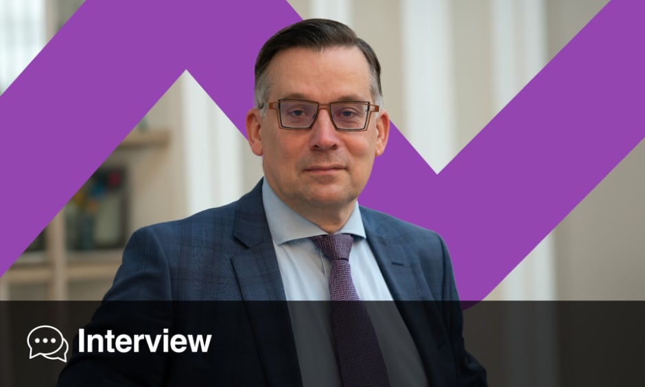
Aad van der Lugt | Professor and Chair Department of Radiology & Nuclear Medicine at Erasmus University Medical Center, Rotterdam, the Netherlands
Citation: EMJ Radiol. 2024;5[1]:40-43. https://doi.org/10.33590/emjradiol/VLXJ1549.
![]()
What was it that inspired you to first begin a career in radiology, specifically neuroradiology?
I have to confess that I did not choose radiology because I found it to be a fantastic job. In the 1980s, there was a labour crisis, and there were actually no available vacancies for residency programmes. My PhD supervisor stepped in and helped me to get a position as a resident in radiology. It took me 2 years to appreciate the profession, but after those initial years, I became committed to radiology for two reasons. The main reason was that I had a hunger for knowledge. Radiology offered an environment with continuous developments in the technical domain. Besides knowledge on imaging physics, we also had to become familiar with various diseases, to interpret the images accurately. In addition, it is not just reporting an exam; you have to know what the relevance is of the abnormalities. In the end, I had to become familiar with two knowledge domains: medicine and physics. So that was, for me, one of the reasons why I truly began to love radiology.
The second reason was that I was exposed to experts in the field, and they were real role models. My supervisor in neuroradiology was consulted frequently to solve difficult cases, and he was so important for the clinical decision-making process. He motivated me to reach the same level in neuroradiology. Thereafter, I was a neuroradiologist, and I was very lucky to have a department chair who was focusing on international development, so he brought in a lot of new trends. He was a great promotor of scientific research and he gave me the opportunity to start my own research programme. I was surrounded by nice, smart people, who actually told me, “come on, try and you can reach the same expert level.” I’ve followed their advice all my life.
You moderated a talk at the European Congress of Radiology (ECR) this year, in Vienna, Austria, on implementing quantitative imaging biomarker protocols in lung cancer screening. Can you give our readers a little bit of information on the European Union (EU)’s lung cancer screening programme decision and implementation plans?
I’m a neuroradiologist, and a head and neck radiologist, so why do I have an interest in lung cancer screening? That’s because I’m Chairman of the European Imaging Biomarker Alliance (EIBALL), which is a subcommittee of the European Society of Radiology (ESR). Our main task is to promote the use of quantitative imaging biomarkers, which are measurements extracted from images, like size, volume, and density. The problem in measuring biomarkers is the variability. When you measure a lung nodule today and tomorrow, it will not have the same size or the same volume, as there is an inherent variability. This variability is increased when you use, for example, a different scan protocol, another scanner, or software. The question arises of how to deal with this variability when the lung cancer screening guidelines propose to do a follow-up exam when the lung nodule size is smaller than 6 mm. EIBALL is dealing with that problem, and trying to solve it.
My main message in the EIBALL session was that, when you start implementing lung cancer screening, you should be aware of the variability, and you should take measures to reduce it, because that will strengthen the study you are performing, and also strengthen the screening programme. We had three presenters: a pulmonologist and a radiologist actively involved in lung cancer screening programmes, and an expert on the imaging biomarker problems. We brought them together and said, “let’s discuss that, and inform the audience about the problems and the solutions.” I was really surprised that the topic was appreciated: the session ran at 8 a.m., and I was little bit afraid that only 10 people would show up. But in the end, it was fully booked!
Could you explain a bit more what EIBALL is, and what its aims are?
Around 10 years ago, in Europe and the USA, we realised that historically, we have used imaging just to diagnose disease. We described the disease, whether it was a round lesion, or an irregular lesion, and we linked these descriptors to biology or outcome. At a certain moment in time, we realised we could also extract measurements: for example the severity of vascular stenosis or the size of the lesion. We felt this was important for radiology and patient care, but we experienced that the measurement had a significant variability, and we realised there was a problem. So, we started trying to quantify the size of the problem. Was it really a problem? Could we reduce the variability in measurements? It would be silly, of course, to do that in every country or professional society; a more harmonised approach would lead to better results. We have collaborated extensively with the USA’s version of EIBALL, the Quantitative Imaging Biomarker Alliance (QIBA), founded by RSNA. We have tried to advocate a common methodology in measuring the variability, and also in solving and reducing variability. I wouldn’t say we are there yet, but progress has been made.
You chaired a session on EIBALL at last year’s ECR. What key developments have taken place in the alliance since then?
So-called metrics have been developed, which help to measure the variability in such a way that you can compare measurements between different sides. Papers have been published on how to do this properly. Of course, this has not fully penetrated the imaging domain yet, but a lot of radiologists and researchers are using these types of metrics. Studies have also been performed to explain how to reduce the variability. With regard to clinical application, studies have been performed that demonstrate that the use of an imaging biomarker is important in the clinical decision-making process. For example, in lung cancer screening, it is clear that you need the measurement of lung nodule size to make a decision about what to do, and that is already a step forward. So, we are trying to increase the evidence base for the use of imaging biomarkers. It is possible to extract multiple biomarkers from an image, and it is important to find out which one is relevant. Artificial intelligence (AI) is also helping us to have a more precise, or fast, measurement of disease characteristics in the image.
What new developments in the field of radiology are you most excited about at the moment?
That’s a very difficult question. Each of us has a narrow view of their own, and an area of expertise, so having a broad view is difficult. However, surprising new developments pop up in many domains. In the image acquisition domain, we have seen CT scanners with photoncounting detectors becoming an exciting topic in the last 2 years, as they provided an increased resolution, a lower dose, and spectral imaging. New contrast agents have popped up in MRI. Sometimes I have the feeling that the field hasn’t changed for 5 years, and nothing new is on the horizon, and suddenly we see exciting new developments, not realising that companies have been working on a topic for more than 20 years before it has reached the imaging community. In image analysis, you can really see that AI is revolutionising the process, not only in the extraction of quantitative imaging biomarkers, but also in detecting disease, aiding diagnosis, and helping to speed up the workflow. For the moment that is the most exciting development, to be honest. But we should not forget the progress in imaging physics and technology!
Speaking of AI, how is the ever-increasing use of AI influencing your practice, research, and teaching as a radiologist and professor?
I have to admit that AI looks like a promising paradise. But it will take years before we will see the fruits of it, because we are still in the process where everyone is trying to pick low-hanging fruit. As an academic radiologist, I have an inborn critical approach, so I like to see the evidence that AI will result in value for patients and society, especially before we invest money. Of course, there are areas where we see improvements in workflow or diagnostic accuracy, but we can’t expand that yet to all domains in radiology. In the coming years, I think there will be a steady progress of penetration of AI in radiology and nuclear medicine.
What do you hope to see ESR and EIBALL achieve in the coming years?
The ESR serves as an important vehicle for radiological education, enabling us to stay up-to-date. Amid our busy daily routines, finding time to extract information from research papers, journals, and websites can be challenging. With the ECR, ESR makes it easy for us to get a quick overview on the current status of radiology. Furthermore, connecting with colleagues and drawing inspiration from these interactions is essential. ESR is also instrumental in harmonising radiology across Europe. By fostering research, ESR is crucial in providing evidence for the important role of imaging in daily clinical care. I sincerely hope that ESR continues to play this crucial role. For EIBALL, I hope that AI and other innovative techniques will significantly enhance biomarker extraction. Overall, I hope that ESR helps us to strive for a future where diagnostic advancements not only improve patient care, but also make our professional lives more fulfilling and enjoyable.
What do you think are the biggest challenges facing young radiologists starting out in the field today?
Feeling overwhelmed. While I acknowledge my own hunger for knowledge, I recognise that many residents may find themselves in a similar situation, but unsure of where to begin. As a radiologist, I often encounter young colleagues who question their abilities and wonder if they can truly reach higher levels of expertise. My message to them is unequivocal: yes, you can. Being selected for a training programme signifies that others believe in your potential. However, it’s essential to understand that progress doesn’t come without effort. Merely sitting in a room and passively listening won’t suffice. Professional growth demands active participation, mental investment, and time. This approach may seem old-fashioned, but it remains effective. As a physician specialising in imaging, you may forget that your commitment extends beyond technical skills. Patient care should remain at the core. Embrace this commitment, invest in your development, and maintain unwavering focus. It’s not merely a hobby; it’s a fulfilling journey. The satisfaction comes when you find yourself in a pivotal role: people seeking your advice, making decisions based on your insights. That, indeed, is rewarding.


