BACKGROUND AND AIMS
Caesarean section, where a baby is delivered via an incision to the abdomen, is currently the most prevalent surgical procedure among patients aged 15–59 years in the UK.1 Globally, rates of Caesarean section deliveries vary, with an average estimated rate of 21%, higher in less developed countries. This number continues to increase, and is projected to reach 30% by 2032.2
While Caesarean sections are generally considered safe, there are a range of associated complications. With the increasing frequency of Caesarean sections, a corresponding rise in the incidence of complications can be expected. Therefore, it is important to ensure that radiologists can effectively identify the most common complications and their features across different imaging modalities.
The purpose of this pictorial review is to depict the various complications associated with Caesarean sections, and their appearances on different imaging modalities using local cases.
RESULTS
The complications identified in this local review of Caesarean section cases align with the recognised complications documented in the literature.
Acute complications included injury to adjacent organs, bleeding, and haemorrhage, along with the risks associated with anaesthesia, typical of acute complications seen with most surgical procedures. The organs most at risk during a Caesarean section are the bladder, ureters, and bowel, due to their proximity to the uterus. Most of the postoperative collections observed were located anterior to the uterus in the uterovesical space, or wound infections in the subcutaneous tissue.
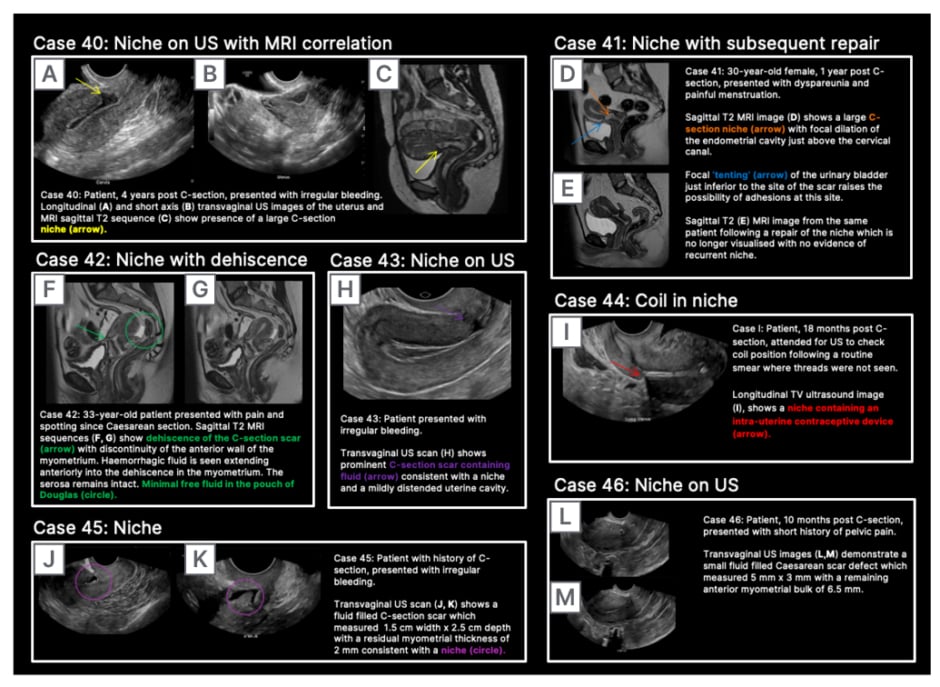
In contrast to acute complications, chronic complications are generally more unique to the Caesarean section itself, and are not typically seen with other surgical procedures, with the exception of incisional hernia. Caesarean scar niche was identified as an important cause of pain and irregular bleeding (Figure 1).

Figure 1: Caesarean scar niche.
C-section: Caesarean section; US: ultrasound.
CONCLUSION
Familiarisation with the complications of Caesarean sections and their imaging appearances is important for radiologists and sonographers in identification and interpretation, and to assist clinicians in the ongoing management of patients.


