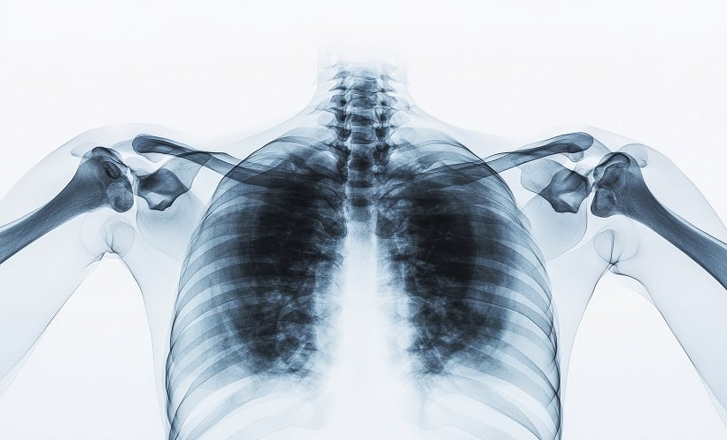
A GROUNDBREAKING study has introduced a novel X-ray detector, designed from silicone elastomer and GOS:Tb, referred to as “imaging skins.” This new technology, developed for integration into custom X-ray systems, converts X-ray radiation into visible light and could play a significant role in surgery, particularly in identifying tumour margins.
The study focused on how different fabrication parameters, such as thickness and concentration, impact sensor performance, especially in real-time clinical applications. Researchers found that varying these parameters did not compromise sensor linearity. The imaging stack demonstrated an exceptional coefficient of determination (R² > 0.99998), indicating that the silicone elastomer did not interfere with the X-ray to light conversion process.
A key finding was the sensor’s ability to stretch, which proved essential for use on curved or irregular surfaces such as human skin or organs during surgery. With a spatial resolution ranging from 1.16 to 1.42 lp/mm, the detector achieved impressive clarity at a thickness of just 0.5 mm.
This study represents a significant step toward the use of stretchable X-ray detectors in clinical environments, offering surgeons an innovative tool for more precise tumour margin detection. However, researchers emphasise the need for further investigation into how the materials interact with X-rays to fully optimise the technology’s potential in complex surgical procedures.
Reference
Dietsch S et al. Image quality evaluation of imaging skins, a novel stretchable X-ray detector for intraoperative tumour imaging. Sci Rep. 2025;15(1):12371.


