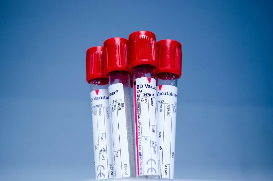
MULTIPLE sclerosis signs can be detected non-invasively with blood samples, according to researchers from the University of Huddersfield, Huddersfield, UK. Advanced mass spectrometry techniques were used to make the discovery, which could also potentially assist with drug development for the condition.
Natural Biomarker Compounds
The team identified sphingosine and dihydrosphingosine, two natural biomarker compounds linked to multiple sclerosis, finding that both were at significantly lower concentrations in blood samples taken from patients with the disease.
Non-Invasive Method
Currently, the detection of multiple sclerosis requires collecting fluid from the brain and spine, a process that is painful and often invasive. This discovery, therefore, provides a far less invasive method of detecting the disease. “Sphingosine and dihydrosphingosine have been previously found to be at lower concentrations in the brain tissue of patients with multiple sclerosis. The detection of these sphingolipids in blood plasma allows the non-invasive monitoring of these and related compounds,” commented the researchers.
New Drug Development
The information from the study was taken using chemometric software, in particular the Mass Profiler Professional (MPP) package. The analysis of compounds associated with multiple sclerosis should improve understanding of their role, and consequently contribute to the development of new drugs. “Mass spectrometry data is very complex and there can be thousands of compounds in each sample,” said co-author Mr Sean Ward, University of Huddersfield. “MPP allows the abundance of each of those compounds to be compared between the samples and can find discrete differences.”
Biomechanical Mechanisms
The investigation of the molecules implicated in multiple sclerosis also enabled an analysis of plasma samples from patients with neuropathic pain, some with and some without multiple sclerosis. Additionally, serum was tested from multiple sclerosis patients who had no neuropathic pain. The researchers were then able to identify the metabolic profiles for each disease state, and the indications were that the three groups share biomechanical mechanisms.
James Coker, Senior Editorial Assistant
For the source, and for further information about the study, click here.


