BACKGROUND AND AIMS
Previous studies support a pivotal role of the pons during the pain phase of a migraine attack.1-3 An altered activation of the pons during trigeminal nociceptive stimulation, as well as resting state functional connectivity (RS FC) alterations between the pons and pain processing cortical areas has been demonstrated in patients with migraine during the interictal phase.4,5 This study aimed to elucidate the role of the pons in migraine pathophysiology using a longitudinal study design and investigate the association between pontine RS FC changes and the clinical characteristics and disease progression of patients with migraine.
MATERIALS AND METHODS
Using a 3.0 Tesla scanner, RS functional MRI and 3D T1-weighted scans were acquired from 91 headache-free patients with episodic migraine and 73 controls at baseline. Twenty-three patients and controls agreed to be re-examined after a median follow-up of 4.5 years. Maps of pontine RS FC were obtained from each subject using a seed-region correlation approach.6 Using a general linear model and SPM12, a whole-brain analysis was performed to assess pontine RS FC changes. The correlation between functional MRI abnormalities and patients’ clinical characteristics was also investigated.
RESULTS
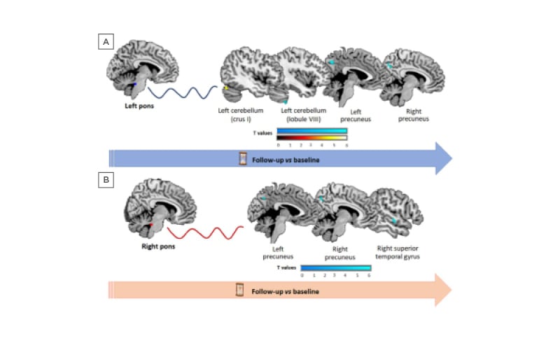
During the follow-up, 11 patients reported a decreased number of migraine attacks, four patients had no changes in migraine attack frequency, and eight patients reported an increased number of attacks. At baseline, compared to controls, migraine patients showed a decreased RS FC between the left pons and left lingual gyrus, right cerebellar lobule V, and bilateral cerebellar crus I; while the right pons had a decreased RS FC with the left cerebellar crus I, right fusiform, and inferior temporal gyrus. Over the follow-up, compared to controls, patients with migraine developed an increased FC between the left pons and the cerebellar crus I, as well as a decreased FC between the left pons, ipsilateral cerebellar lobule VIII, and bilateral precuneus (Figure 1A). Patients with migraine also experienced a decreased right pontine RS FC with the ipsilateral superior temporal gyrus and bilateral precuneus (Figure 1B). The increased RS FC between the left pons and ipsilateral cerebellar crus I was associated to an increased migraine attack frequency over the years.

Figure 1: Brain regions showing significant longitudinal resting state functional connectivity changes of the left (A) and right (B) pons in patients with migraine compared to controls at follow-up versus baseline.
Areas of decreased RS FC are colour-coded in blue, and areas of increased RS FC are represented in red, according to their t values. The left pons is represented in blue, and the right pons is represented in red. RS FC: resting state functional connectivity; vs: versus.
CONCLUSION
Similar to previous cross-sectional studies,4,5 the authors confirmed that the pons modulates the activity of pain processing brain areas in patients with migraine. Their findings also highlighted an interaction between the pons and extrastriate visual areas during the interictal phase, supporting their key role in migraine pathophysiology.
In this cohort, the RS FC of the pons changes dynamically over time. After 4 years, the RS FC between the pons and precuneus, a region known to be involved in sensory integration, is decreased. Compared to controls, patients with migraine developed both decreased and increased RS FC between the pons and the cerebellum, suggesting that distinct adaptive responses might occur over time at the level of the cerebellum. Some of the cerebellar changes might represent a maladaptive response, contributing to migraines worsening.


