Calcium signalling is essential for inter and intra-cellular neuronal communication, and efforts for improving its imaging are constantly ongoing due to the insights it would provide into neurological disorders. A significant hurdle so far has been the depth to which we can generate images; however, a team from Massachusetts Institute of Technology (MIT), Cambridge, Massachusetts, USA believes they have made a breakthrough.
When neurons are electrically active, their calcium concentration massively increases, and to date this has been detected by labelling the calcium using fluorescent molecules. This can be done both in vitro and in vivo, but as this type of fluorescence microscopy can only work at a depth of a few tenths of a millimeter, visualising the electrical activity in the interior of the brain was impossible.
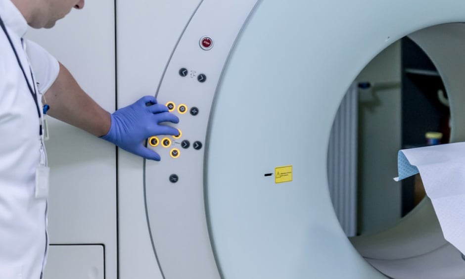
Now, MIT researchers have developed a technique using MRI to bypass this problem. They have synthesised a compound containing manganese that is bound to an organic compound capable of permeating cell membranes, as well as a calcium-binding chelator. When calcium is absent, the signal from the manganese is masked due to being weakly bound to the calcium chelator and the signal is diminished; however, in instances where calcium intake is high (i.e., electrically charged neurons), the manganese is unbound and produces a brighter contrast during the MRI scan.
The team were able to demonstrate the method’s effectiveness in rats, successfully measuring calcium dynamics with their sensor following stimulation with potassium ions. Temporal and spatial neuronal activity can now be measured deep within the brain, looking specifically at sub-sets with defined functions.
Senior author Prof Alan Jasanoff, MIT, comments: ‘‘This could be useful for figuring out how different structures in the brain work together to process stimuli or co-ordinate behaviour.’’
Beyond the neurological applications, the group speculates that the technique could even be used for diagnostic purposes in other organs that rely on calcium, such as the heart, as well as for analysing calcium function in other processes, such as immune cell activation.


