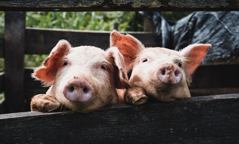
HEPATOCYTES are natural regenerators, but they require a nurturing environment to multiply. Researchers at the University of Pittsburgh School of Medicine, Pittsburgh, Pennsylvania, USA, have found that pig lymph nodes can provide such an environment, with the capacity to restore a degenerated liver.
Almost 10 years ago, Prof Eric Lagasse, senior author of the study, discovered that injecting healthy liver cells into the lymph nodes of a mouse with a genetically-induced malfunctioning liver led to the formation of an auxiliary liver that was able to take over liver functioning. He explained: “If hepatocytes get in the right spot and there is a need for liver functions, they will form an ectopic liver in the lymph node.”
However, Prof Lagasse wanted to know if this related to larger animals too. By diverting the main blood supply away from the liver and simultaneously injecting hepatocytes, which had been extracted from the pig’s healthy liver tissue, into the lymph nodes, the researchers found that all six pigs had recovery of function. Examination of the new lymph nodes, revealed thriving hepatocytes and a network of bile ducts and vasculature that had spontaneously formed.
Additionally, the size of the auxiliary liver was proportional to the severity of liver dysfunction; the more damaged the liver, the bigger the supporting liver. “It’s all about location, location, location,” said Prof Lagasse.
These results are supplementary to another recent study by Prof Lagasse and his colleagues, whereby they found that healthy liver tissue grown in the lymph nodes of pigs with a genetic liver defect spontaneously migrated to the liver. The healthy tissue was then able to reinstate the dysfunctional cells and the animal was cured of the disease.


