CYSTIC echinococcosis (CE) is a severe zoonotic parasitic disease caused by the larval stage of Echinococcus granulosus, a tapeworm primarily affecting the liver. CE presents a significant global health challenge, impacting over one million people worldwide and incurring an estimated annual healthcare cost of $3 billion. The WHO has classified echinococcosis as one of the 17 neglected tropical diseases targeted for control or elimination by 2050.
CE is particularly prevalent in regions with intensive livestock farming, including Central Asia, South America, and the Middle East. Incidence rates vary widely, from 1 to 200 cases per 100,000 individuals. The liver is the most commonly affected organ, accounting for over 70% of cases, with a mortality rate between 2% and 4%. The disease’s geographic distribution has expanded due to globalization, increased population mobility, and evolving pet ownership patterns. Despite advancements in diagnostic imaging, CE remains underreported in many regions, and its diverse imaging manifestations pose significant diagnostic challenges.
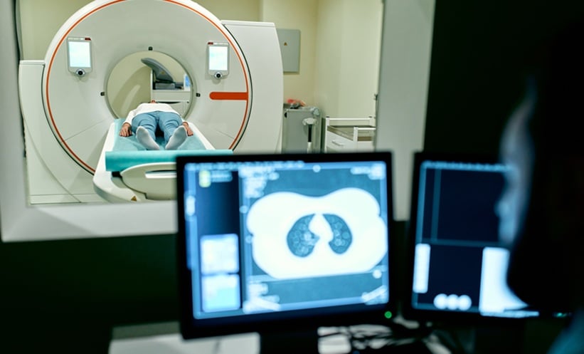
Imaging modalities such as ultrasound, CT, and MRI play vital roles in diagnosing and managing hepatic CE. These techniques provide crucial details about cyst location, morphology, and characteristics, each offering unique strengths in lesion characterization. CT, for example, excels in spatial resolution and density assessment, making it a valuable diagnostic tool.
A recent study analysed CT imaging findings from 48 pathologically confirmed cases of hepatic CE across two medical centers. The study aimed to systematically categorise CT imaging manifestations, correlate imaging features with pathological findings, and develop a CT-based diagnostic algorithm. This approach enhances diagnostic accuracy and streamlines evaluation in regions where CE is emerging or underrecognized. The study findings suggest that the pathological progression of CE is well reflected in imaging results, particularly regarding cyst wall structure, internal architecture, and calcification patterns. The CT diagnostic algorithm demonstrated 94% accuracy in classification and high inter-observer agreement.
While CT provides detailed morphological information, other imaging modalities contribute additional benefits. Ultrasound remains the first-line examination in endemic regions due to its affordability and lack of radiation exposure. MRI excels in soft tissue contrast, while PET scans assess metabolic activity. Combining these imaging techniques enhances diagnostic precision and treatment planning. Future research should focus on optimizing imaging-based diagnostic criteria and evaluating long-term outcomes to improve CE management.
Katie Wright, EMJ
Reference
Zhang H et al. CT imaging features and diagnostic algorithm for hepatic cystic echinococcosis. Sci Rep. 2025;15(1):10671.


