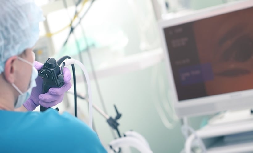
A NEW study presented at the United European Gastroenterology (UEG) Week 2024 is exploring a breakthrough imaging technique aimed at improving treatment outcomes for patients with locally advanced oesophageal cancer (EC).
Currently, patients with EC are treated with a combination of neoadjuvant chemo(radio)therapy (nCRT) followed by surgery, but only 16–43% achieve a complete pathological response. To improve these odds, researchers are investigating the use of immune checkpoint inhibitors (ICI) that target PD-1 and PD-L1 proteins, but effective patient selection for ICI therapy remains challenging.
This new study tests a novel imaging method, ultrasound-guided quantitative fluorescence molecular endoscopy (US-qFME), designed to visualise PD-L1 protein levels in tumours. By using the fluorescently labelled ICI drug durvalumab-680LT, the technique allows for the visualisation of PD-L1 expression, offering a way to better select patients for ICI therapy. The aim is to assess both the safety and effectiveness of the technique in monitoring drug distribution across cancerous tissues before and after nCRT.
Fifteen EC patients scheduled for nCRT have been enrolled in the study so far, divided into different dose groups receiving 4.5 mg or 15 mg of durvalumab-680LT, or no dose as a control. A final group of patients will receive 25 mg of the drug.
Results so far are promising: fluorescence signal intensities were higher in tumour biopsies compared to healthy oesophageal tissue in both the 4.5 mg and 15 mg dose cohorts, indicating that durvalumab-680LT successfully targeted tumour tissue. Mucosal and ultrasound-guided spectroscopy gave similar results, with higher signals in tumour tissue both in vivo and ex vivo compared to healthy tissue. Furthermore, a broad range of fluorescence signals were observed within tumours, suggesting variability in PD-L1 expression among patients.
So far, the procedure has been well-tolerated, with no serious adverse effects reported. Further analysis will determine how well this technique correlates with traditional PD-L1 staining methods, and its potential to improve patient selection for ICI treatment. If successful, this innovative approach could guide more effective, personalised treatments for patients with EC in the future.
Ada Enesco, EMJ
Reference
Van der Waaij AM et al. Optical PD-L1 imaging using ultrasound-guided quantitative fluorescence molecular endoscopy combined with durvalumab-680LT in locally advanced esophageal cancer patients. OP065. UEG Week 2024, 12–15 October, 2024.


