Abstract
The specific dietary intervention known as exclusive enteral nutrition (EEN) is well-established as the preferred treatment to induce remission in children with active Crohn’s disease. The majority of children managed with EEN respond well to this intervention, with high rates of mucosal healing, improved nutrition, and enhanced bone health, with few side effects. This dietary therapy, utilising a complete nutritional liquid product, is generally well-tolerated over the short period of induction of remission, but does require substantial changes to routine oral intake and daily patterns. After a period of exclusive use of this therapy, ongoing use of the same formulae (as maintenance enteral nutrition) may prolong remission and prevent relapse. Over the last few years, new reports have advanced our understanding of the mechanisms by which EEN acts: these include modulation of the intestinal microbiota and direct anti-inflammatory effects upon the epithelium. This review highlights key outcomes of EEN in children with Crohn’s disease and highlights the current understanding of the mechanisms of action.
INTRODUCTION
The inflammatory bowel diseases (IBD) are a group of conditions characterised by chronic, incurable inflammation in the gastrointestinal (GI) tract.1,2 The diagnosis of IBD is based upon endoscopic and histologic features, along with altered inflammatory markers, and radiology results.1 The two main classifications of IBD are termed Crohn’s disease (CD) and ulcerative colitis. These conditions are generally defined by their location in the gut, the pattern of inflammation, and disease behaviour.
At present the exact cause of IBD is not known definitively. The currently most-accepted hypothesis is that IBD begins in an individual with genetic risk (>240 genes are now linked to IBD) when various environmental factors trigger changes in the intestinal microbiota, prompting innate and acquired immune responses that are then dysregulated.3-6 The role of genetic factors may be more pronounced in younger children than in adults; this is paramount in the increased identification of monogenic forms of gut inflammation, as seen in children aged <6 years of age (very-early onset IBD).
Given the incurable nature of IBD, the focus of management is firstly upon the induction of remission, and then on the subsequent maintenance of remission. Establishment of mucosal healing is identified as a key outcome of management, with pronounced impact upon the subsequent disease course. Nutritional interventions, especially exclusive enteral nutrition (EEN), provide an important and safe method to induce remission and establish mucosal healing, particularly in children with CD. This review aims to outline the key aspects of CD, with a particular focus on children, and to overview the role EEN can play in children with CD, highlighting the putative mechanisms of this nutritional intervention.
CROHN’S DISEASE
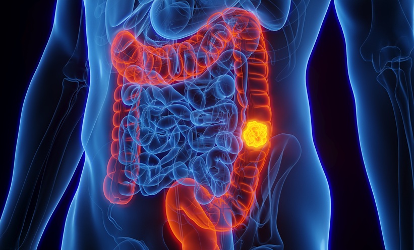
CD is characterised by the presence of discontinuous inflammatory changes in any section of the GI tract.1,2 Inflammation is typically transmural, with disease defining features of skip lesions and non-caseating granulomata. CD begins with an inflammatory phenotype which can then be complicated by the development of either fistulising (penetrating) or stricturing disease. Although some children will present with disease complications, most will have purely inflammatory luminal disease at diagnosis.7,8 CD can also be accompanied by the presence of various extra-intestinal manifestations, which include peri-oral or oral findings, joint disease, eye changes, or skin manifestations.9
Children can be diagnosed with CD at any age, but it is more common in the second decade.1 Although typical symptoms include the combination of weight loss, diarrhoea, and abdominal pain, other children may have atypical symptoms such as isolated linear growth failure, or weight loss without associated GI symptoms. Atypical symptoms may impede the recognition of CD, leading to diagnostic delay.
Almost all children diagnosed with CD will have weight loss, or impeded weight gains, which is mostly mediated by early satiety, post-prandial pain, or diarrhoea and consequent reduced dietary intake.10,11 In addition, malabsorption may contribute. The anorexic effects of circulating inflammatory mediators, such as TNF-α, contribute to these outcomes. Reduced dietary intake and altered weight gains may then result in impaired linear growth, especially in peri-pubertal children, due to interrupted pubertal growth spurt.12 A further consequence of active CD is delayed onset of puberty: this is more commonly observed in adolescent boys. These nutritional changes may reflect disease severity and may lead to significant psychological adverse effects. Furthermore, interruption of normal adolescent developmental processes may lead to reduced final adult height.
In addition to the adverse impacts upon nutrition and growth in children diagnosed with CD, the patterns of CD present in children also differ in other regards from the same disease presenting in adult years. As illustrated in two large cohorts from France and Scotland, paediatric CD is typically more severe and extensive, with pan-enteric disease distribution seen commonly.7,8 For example, both cohorts described higher rates of upper gut involvement in children with CD.7,13
EPIDEMIOLOGY OF CROHN’S DISEASE
Numerous studies have shown increasing rates of IBD in the last few decades.14 From around the start of the 21st century, there have been particular increases in diagnoses in Asian countries.15 In addition to these broad changes in IBD patterns, there have been continued increases noted in children and adolescents; for example, high rates of IBD (and especially CD) were noted in 2004 in the Canterbury region of New Zealand (NZ).16 A subsequent study 10 years later showed an increase of almost two-fold within the same region.17 Furthermore, a longer-term study focussing on children diagnosed in the same region of NZ demonstrated almost a five-fold increase in incidence over two decades until 2015.18 In addition, high prevalence was also demonstrated in another study that determined the numbers of children diagnosed across the entirety of NZ.19
The reasons for high rates of IBD in various parts of the world, including NZ, and for the recent increased incidence, are unclear. Environmental factors are likely the most important drivers of these changes.20 While vitamin D and sunlight exposure appear to explain some regional differences (with increasing rates with increasing latitude), dietary factors appear most important. Several reports indicate that breastfeeding and childhood pet ownership are protective. Westernised diets (high fat, high sugar foods), urbanisation, and dietary additives or preservatives are also implicated in higher rates of IBD.20 Other early life events (for example birth method and antibiotic exposure) may further contribute to increased risk.
THE NUTRITIONAL IMPACT OF CROHN’S DISEASE IN CHILDREN
Many children with CD have a history of weight loss or poor weight gains prior to diagnosis. Some will also have impaired linear growth and others may have delayed pubertal development.1,10,11 Micronutrient deficiencies can also occur in children with CD.
Poor weight gains are most commonly secondary to decreased oral intake, with early satiety and pain limiting intake. The circulating pro-inflammatory cytokines, such as IL-6, have been shown to induce anorexia, which contributes to these changes. While poor diet and weight gains may have a role, impaired linear growth is primarily a result of uncontrolled inflammation, including elevated IL-6, resulting in lower production of insulin growth factor 1 (IGF-1) and related proteins, which in turn abrogate the effects of growth hormone.12
Further to the adverse effects of reduced caloric intake and macronutrients, micronutrient deficiencies are also common in children with CD.10,21-23 While low levels of iron and vitamin D are seen most often, zinc, selenium, and vitamin B12 may also be low. Low iron stores, consequent to reduced intake, impaired absorption, or increased enteric losses, result in anaemia, and present as fatigue, lethargy, and disrupted learning in children. Numerous reports indicate that children with CD typically have lower vitamin D levels than control children.24,25 In an Australian report, more than half of a group of 78 children were deficient at diagnosis.24 Vitamin D is critical for bone health but also contributes to innate immune function.26 Correction of vitamin D levels has been associated with reduced inflammatory activity;27,28 however, the ideal required level is unknown.
In view of the various nutritional impacts of CD in children, this needs to be a central aspect of management goals, in which nutritional therapies play a critical role.
EXCLUSIVE ENTERAL NUTRITION
A Typical Exclusive Enteral Nutrition Protocol
EEN involves the use of a liquid formula providing all nutritional requirements for a defined period, commonly 8 weeks, along with exclusion of usual solid foods.29,30 Case reports and series published more than three decades ago described reduced inflammatory activity in adults taking intensive nutritional supplementation.31-35 These observations were supported in an Irish randomised controlled trial that showed that EEN had similar outcomes to corticosteroids.36 In more recent years, a large body of data has demonstrated that EEN has tremendous benefits to children with CD,30,37 such that it is now recommended by European and North American organisations as the best therapy to induce remission in a child with active CD.38-40 However, the utilisation of EEN and the specific EEN regimens vary between regions and countries.41,42
EEN is generally well-tolerated with few side effects expected. Refeeding syndrome has been reported in a handful of cases.43,44 Although one group showed transient elevation of serum transaminases during EEN, this finding was not replicated in a second report.45,46
Exclusive Enteral Nutrition and Induction of Remission
The primary role of EEN is the induction of remission in children with active CD, especially at diagnosis. Numerous paediatric reports and meta-analyses of paediatric data clearly show that EEN has similar efficacy to corticosteroids;47 however, not only does EEN avoid steroid-related side effects, which include impaired linear growth, EEN also leads to enhanced rates of mucosal healing, which is a key treatment target.48 EEN appears to have optimal benefits at the time of diagnosis,49 with lower remission rates in those with long-standing disease. Generally, paediatric studies indicate remission rates of 80–85% with mucosal healing seen in up to 75% of those entering remission.30,39
A recent meta-analysis focussing on the outcomes of EEN in children with CD did not delineate any difference in efficacy to that seen in children managed with corticosteroids.37 This report included 18 studies, 4 of which were prospective randomised controlled trials. Although efficacy between the two interventions was found to be similar, EEN resulted in much greater rates of endoscopic mucosal healing (odds ratio: 5.4; p=0.0005) and histological healing (odds ratio: 4.78; p=0.0009). Furthermore, weight gain was greater with EEN.37
As a further assessment of response to EEN, a number of earlier publications have evaluated various stool-based noninvasive markers such as calprotectin, S100A12, and osetoprotegerin during and following EEN in children.50-52 Gerasimidis et al.52 showed that faecal calprotectin decreased in children who entered clinical remission during EEN, but levels fell to the normal range in only one child. The level of reduction at 30 days correlated with response at the end of the EEN course. In contrast, Copova et al.53 did not demonstrate any association between early reduction in calprotectin at 2 weeks and clinical response at 6 weeks. More recently, Logan et al.54 reported that the reduction in calprotectin seen during EEN was not maintained after the recommencement of standard solid diet at the end of the EEN course.
Nutritional Benefits of Exclusive Enteral Nutrition
EEN also has benefits on nutritional status, including growth parameters, micronutrients, and bone health. Weight gain is expected during a course of EEN, especially in those with malnutrition prior to diagnosis.30,39 The impact of EEN upon linear growth is less clear. One retrospective report showed that height increments over 24 months from diagnosis were greater (p=0.01) in 31 children treated with EEN than in 26 children managed with corticosteroids.55 In contrast, a more recent report evaluating height outcomes 18 months after diagnosis in an inception cohort of Canadian children did not show any difference between those managed with EEN or corticosteroids.56
Nutritional changes occur early after starting EEN, as illustrated by prompt increases in markers such as IGF-1.57,58 Children with active CD have altered bone health (reduced new bone formation and increased breakdown) compared to their age-matched peers. In an Australian study, bone health improved within 6–8 weeks of EEN, with enhanced new bone formation and reduced breakdown evident.59 Other reports indicate improved bone mineral density in children with CD after treatment with EEN.60
Ongoing Enteral Nutrition to Maintain Remission
After induction of remission with EEN, some reports indicate benefit from ongoing use of enteral nutrition in conjunction with normal diet to maintain remission (i.e., maintenance enteral nutrition). This strategy may also work well in combination with medical therapies, enhance growth, and prevent relapse after surgically induced remission.
Early studies conducted in Canada showed that intermittent periods of EEN (such as given for 1 month every 3 months) or overnight feeds (given in conjunction with normal diet during the day) resulted in prolonged remission.61,62 Remission may also be maintained with the addition of day-time sip feeds along with normal diet.63 The ideal volume and caloric intake delivered in this fashion is not clear.
Further, a recent Scottish report noted that a small group of children who received minimal enteral nutrition were able to maintain lower levels of faecal calprotectin, suggesting enhanced mucosal control.54 Despite this, minimal enteral nutrition in this group of 15 children was not associated with longer duration of remission.
Several Japanese studies have shown that on going feeds given overnight prevents disease relapse. In one of these reports, a group who received half their recommended caloric intake as an elemental feed overnight were half as likely to relapse than a comparative group who did not receive overnight feeds.64 Other studies from Japan have demonstrated lower rates of recurrence after surgical resection with maintenance nutrition.65-68
Recent reports have shown that nutritional support may work in concert with biologic therapies to augment response and prevent secondary loss of response.69,70
Given that EEN and anti-TNF-α inhibitors are the most effective interventions to result in mucosal healing, further work on such combined regimens may lead to important enhanced outcomes.71-73
Exclusive Enteral Nutrition and Complicated Crohn’s Disease
Studies evaluating EEN have typically focussed on individuals with inflammatory CD alone. However, a number of reports have indicated that EEN may also have a role in the management of patients with complicated CD (penetrating or stricturing disease).
Two earlier publications described the inclusion of EEN in the management of a teenager with an entero-vesical fistula and three children with fistulising perianal disease.74,75 More recently, EEN was beneficial in the management of two teenagers who presented with an ileal fistula and associated collection (phlegmon).76 After an initial short period of gut rest, parenteral nutrition, and antibiotics, both children entered remission with an uncomplicated period of EEN. EEN may also be helpful in other manifestations of CD: for instance, 1 group reported that 19 out of 22 children with peri-oral changes had improvement after 8 weeks of EEN.77
This paediatric experience has been followed by a number of reports of EEN having a role in adults for the management of internal fistula with phlegmon, enterocutaneous fistula, and stenotic disease.78-81 For example, a report from China described that 12 weeks of EEN resulted in healing of enterocutaneous fistulae in 30 out of 48 adults treated with EEN alone for 12 weeks.82
MECHANISMS OF ACTION OF EXCLUSIVE ENTERAL NUTRITION
A number of publications in the last decade have focussed upon the mechanisms of action of EEN in CD.83 While gut rest and avoidance of one or more dietary triggers has been considered in the past, the more recent findings indicate that active effects of EEN are likely more important. Overall, this work has focussed on the effects of EEN upon the intestinal microbiota, improved barrier function, increased production of innate defence proteins, and direct anti-inflammatory activity.
Many reports have clearly shown that substantial changes are seen in the intestinal microbiota during and following the administration of EEN.84-91 The application of advanced molecular tools such as 16S rRNA high-throughput sequencing and whole genome or shot-gun sequencing, have enabled researchers to show early and profound alterations in the microbiota. For example, one of these publications examined the flora before EEN, after 2 weeks, and then at the completion of EEN in 15 children with active CD.90 The proportion of bacteria belonging to the Bacteroidetes phylum reduced while those in the Firmicutes phylum were increased. Further to the changes in the intestinal microbiota, this article showed concurrent changes in regulatory T lymphocytes. Although the authors did not delineate the exact connection between these separate processes, they did propose that these events may also contribute to the benefits of EEN.
Another recent report on the intestinal microbiota changes consequent to EEN demonstrated that initial changes in microbial diversity were linked with the outcome of a subsequent sustained remission.91 This finding was used to predict the outcome with 80% accuracy.
These paediatric studies are complemented by two studies examining alterations in diversity in adults managed with EEN: both showed similar patterns.92,93 In addition to the various reports that have focussed on the intestinal microbiota after EEN in individuals with CD, one report has evaluated the impact of EEN in children with rheumatologic disease managed successfully with EEN.94 Similar changes in the microbiota were shown, suggesting that the alteration in the bacterial patterns reflect the dietary change. The precise reasons that these events abrogate inflammation have not yet been elucidated.
A series of in vitro and animal studies has demonstrated that the polymeric formulae (PF) used for EEN generates several changes in key components of intestinal barrier function. Complementary evaluations of barrier function (such as trans-epithelial electrical resistance, short circuit current, para-cellular permeability, and tight junction protein patterns) in an epithelial cell line model of gut inflammation demonstrated that PF reversed the detrimental effects of pro-inflammatory cytokines.95 Furthermore, these corrections of intestinal tight junction activity were mediated by inhibition of myosin light-chain kinase. Experiments conducted using an animal model of colitis showed consistent findings.
Further to these data, other reports using intestinal epithelial cell lines and animal models of IBD show direct anti-inflammatory effects consequent to PF.96,97 These appeared to be modulated by interruption of NF-κB signalling. Further work demonstrated that these effects were mediated by arginine, glutamine, and vitamin D3; all present within the PF. Glutamine and arginine directly modulated components of the NF-κB and p-38 signalling pathways.98
In addition, recent work has shown that PF leads to carcinoembryonic antigen-related cell adhesion molecule (CEACAM)-6 and intestinal alkaline phosphate, two separate innate defence proteins.99,100 The first of these reports demonstrated that PF exposure of epithelial cells resulted in increased production of CEACAM-6, which then functioned as a soluble decoy by binding bacteria thereby preventing bacterial interactions with the epithelial cells.99
While these reports have focussed on models of gut inflammation and do not include human studies, they do provide important and consistent support for the direct anti-inflammatory effects of EEN. In addition, the relationship(s) between these separate findings have not yet been ascertained.
CONCLUSION
EEN is recommended in several international guidelines as the preferred primary therapy for the induction of remission in children with active CD. These recommendations are based on the evidence that this therapy is safe and effective in children and adolescents.
Although there have been numerous studies focussing on the mechanisms of EEN, the precise mechanism of action has not yet been fully ascertained. It is most likely that these effects are direct and do not just reflect gut rest. In addition, it also appears feasible that the effects of EEN upon the intestinal microbiota might mediate some of the observed anti-inflammatory effects.
While the benefits of EEN are clear and unquestionable, EEN does not cure CD. In addition, EEN is not feasible to maintain indefinitely due to the onerous requirements to avoid solid food and the consequent disruption of normal dietary habits. Other interventions are consequently needed to maintain remission and prevent relapse. Maintenance enteral nutrition is one potential way to achieve this, but the optimal regimen to undertake this on an ongoing basis has not been demonstrated. Optimisation of EEN regimens, with enhanced provision of active components such as glutamine, may be beneficial.
Despite these reservations, it is clear that EEN has a role in switching off the inflammation seen in CD. Further understanding of the mechanisms of these events may also provide clues to the aetiology of CD.


