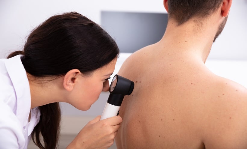
A CLINICAL trial has assessed whether 3D total-body photography (TBP) combined with sequential digital dermoscopy imaging (SDDI) enhances melanoma detection in high-risk patients. Conducted at a research hospital in Brisbane, Australia, from 2018 to 2021, the study compared usual clinical care with an intervention group receiving additional 3D TBP and SDDI every six months via teledermatology.
The study included 314 participants (mean age 51.6 years, 62% female), with 158 in the intervention group and 156 in the control group. Over the study period, a total of 1,527 excisions were performed, 905 in the intervention group and 622 in the control group. Despite the higher number of biopsies, fewer melanomas were detected in the intervention group (24 cases, 35%) compared to the control (43 cases, 64%). The incidence rate of melanoma per person was lower in the intervention group (0.08 vs. 0.16), but this difference was not statistically significant.
While 3D TBP led to a greater number of skin excisions and biopsies, its impact on melanoma detection remains unclear. Researchers emphasize the need for further studies comparing teledermatology with standard care and exploring the potential of AI to enhance diagnostic accuracy.
This study highlights the feasibility of integrating teledermatology into melanoma surveillance, though larger trials with diverse patient populations and more teledermatologists are needed to determine its clinical value.
Reference: Soyer HP et al. 3D Total-Body Photography in Patients at High Risk for Melanoma: A Randomized Clinical Trial. JAMA Dermatol. 2025:e250211. doi: 10.1001/jamadermatol.2025.0211. [Online ahead of print].
Anaya Malik | AMJ