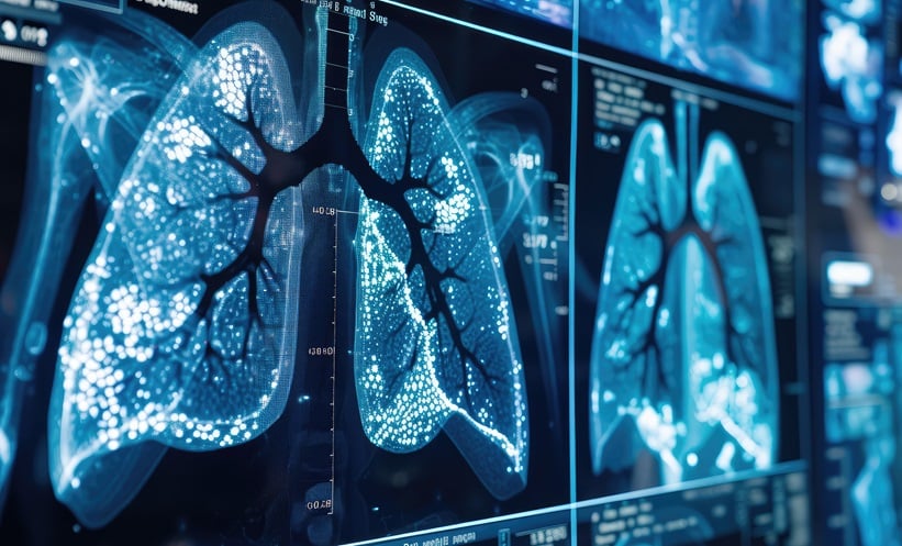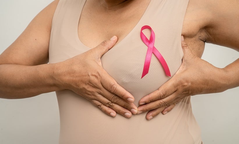
BREAST MRI is widely recognised for its sensitivity in detecting small breast cancers. Despite this, distinguishing between benign and malignant lesions remains a challenge, particularly for foci, which are often difficult to interpret. A recent study aimed to determine whether the kinetic features of ultrafast MRI (UFMRI) could aid in differentiating malignant from nonmalignant foci. A key finding was that the difference in time to enhancement (TTE) between a lesion and the surrounding background parenchymal enhancement (BPE) could be a significant indicator of malignancy.
The retrospective analysis was conducted at a single centre, spanning from July 2019 to April 2023. It focused on patients who had undergone UFMRI examinations and subsequently had MRI-guided biopsies of foci. Key data was gathered from electronic medical records (EMR), imaging reports, and reviews of the imaging. The main variables included the TTE for both the lesion and BPE, as well as the difference in these times, which was analysed for its association with malignancy through univariable and multivariable logistic regression.
A total of 124 patients, with a mean age of 53, were included in the analysis. The results revealed that 21 (16.9%) of the foci were malignant, including 16 invasive cancers and five cases of ductal carcinoma in situ. The odds of malignancy increased by 5% for each 1-second increase in the difference between the lesion TTE and BPE TTE (95% CI: 2–9%; p=0.006). Additionally, older age and lower BPE were associated with a higher likelihood of malignancy. Notably, patients with minimal or mild BPE had 11.69 times the odds of malignancy compared to those with moderate or marked BPE (95% CI: 1.51–90.67). These findings indicate that kinetic characteristics on UFMRI may be a useful tool for identifying malignancy, potentially reducing unnecessary biopsies.
In conclusion, the study provides evidence that earlier visualization of a lesion relative to BPE on UFMRI is associated with an increased likelihood of malignancy. This kinetic parameter could aid in clinical practice by assisting in the identification of malignant lesions, thereby helping to reduce unnecessary biopsies. However, the study is limited by its single-centre design and retrospective nature, which may limit the generalisability of the results. Further multicentre prospective studies would be beneficial to validate these findings and confirm their broader applicability in clinical settings.
Reference
Regen-Tuero HC et al. Time to Enhancement of Foci Relative to Background Parenchymal Enhancement on Ultrafast Breast MRI: A Single-Center Retrospective Study. AJR Am J Roentgenol. 2025;DOI: 10.2214/AJR.24.32309.