OBJECTIVE
To explore the spectrum of MRI abnormalities in patients with rabies encephalitis.
BACKGROUND
Rabies is a highly lethal infectious disease and is equivalent to a death sentence for patients. The ante-mortem diagnostic sensitivity is only approximately 40% with currently available tests. Currently, there are only isolated case reports1 or small case series exploring the MRI features in rabies encephalitis.
METHODS
A prospective observational study was designed to explore the patterns of MRI abnormalities in rabies patients. A total of 31 patients were enrolled in the study, out of which 21 underwent contrast enhanced-MRI of the brain and spinal cord.
RESULTS
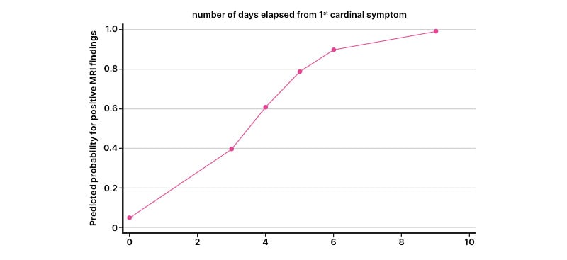
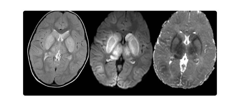
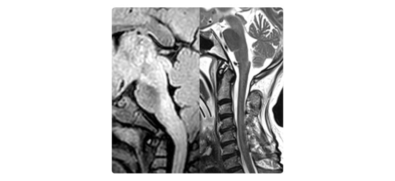
Overall, MRI was normal in 10 (47.6%) patients, whereas 11 (52.4%) patients showed abnormalities. It was observed that those with abnormal MRI tended to have longer time to MRI from symptom onset as compared to those whose MRIs were normal (mean number of days: 4.5 days versus 2.9 days; Figure 1). In addition, univariate logistic regression analysis revealed that the likelihood of picking up abnormalities on MRI increases with the duration from the symptom onset, approaching approximately 80% at Day 6 and >90% at Day 8. Abnormalities were detected in various brain regions, with brainstem, spinal cord and basal ganglia being the most affected regions (Figure 2 and 3). Other regions such as the cortex, sub-cortical white matter, limbic system, internal capsule, corona radiata, thalamus, and sub-thalamic region were also involved to varying degrees. Diffusion-weighted-imaging showed a typical pattern of diffusion restriction in six cases (28.5%). No gadolinium contrast enhancement was observed in any patient in the affected brain areas; however, some patients demonstrated contrast enhancement in the spinal nerve roots. Overall, no significant difference in neuroimaging findings were observed between encephalitic and paralytic forms of rabies.

Figure 1: Predicted probability of MRI being abnormal based on the number of days elapsed from the onset of first cardinal symptom.

Figure 2: T2 turbo spin echo showing hyperintensities in bilateral basal ganglia (caudate nucleus, putamen, and globus pallidus) and thalamus and corresponding diffusion-weighted imaging and apparent diffusion coefficient images showing true diffusion restriction.

Figure 3: Fluid-attenuated inversion recovery/T2 hyperintensities in dorsal brainstem and cervical spinal cord of two different patients.
Diffusion-weighted-imaging showed a typical pattern of diffusion restriction in six cases (28.5%). No gadolinium contrast enhancement was observed in any patient in the affected brain areas; however, some patients demonstrated contrast enhancement in the spinal nerve roots. Overall, no significant difference in neuroimaging findings were observed between encephalitic and paralytic forms of rabies.
CONCLUSION
To date this is the largest prospective study (21 cases) exploring the MRI findings patients with rabies encephalitis. The study demonstrated certain patterns of brain imaging in rabies that, in the appropriate clinical context, could significantly aid in establishing the ante-mortem diagnosis of rabies encephalitis. At the same time, the study also highlighted the fact that a normal MRI brain does not rule out the diagnosis and that the probability of finding specific abnormalities on MRI increases as the disease progresses.


