BACKGROUND
Rabies is the deadliest infectious disease, with a nearly 100% fatality.1 Due to this fear, it is often an overlooked disease, resulting in a scarcity of comprehensive knowledge, most of which is derived from retrospective studies.2,3 This prospective study is aimed to narrow this knowledge gap.
METHODS
This prospective observational study included 31 participants. Detailed clinical history and clinical examination were performed, and the Milwaukee protocol was used for patient management. For diagnostics, CEMRI Brain was performed, and samples of cerebrospinal fluid, saliva, serum, skin, and brain biopsies were collected for various diagnostic tests. An autopsy was done for eight patients.
RESULTS
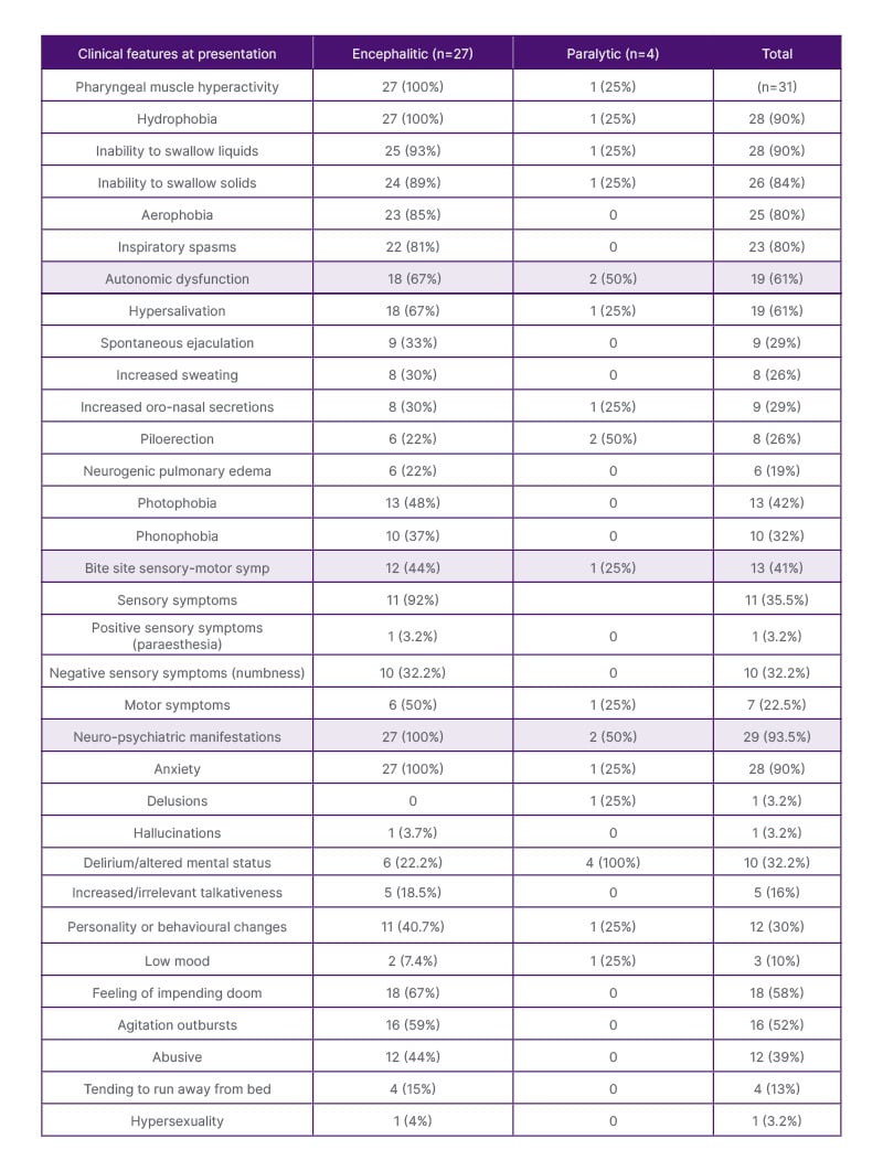
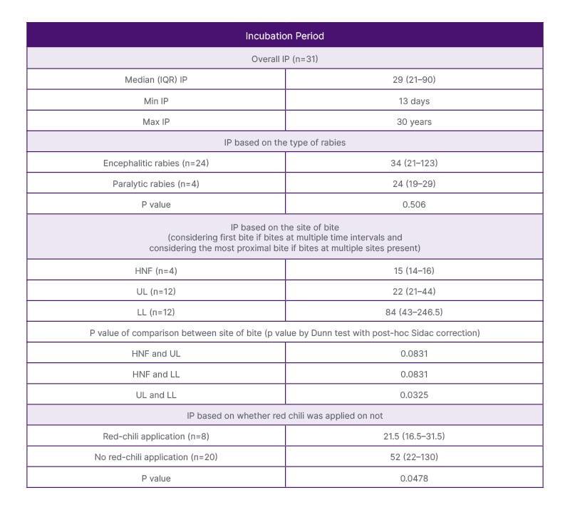
The study unearthed numerous intriguing aspects of the disease. Despite the presence of adequate infrastructure, only 48% initiated post-exposure prophylaxis, and a mere 13% received Rabies immunoglobulin. Adherence to the vaccination schedule was subpar, with only 13.3% completing the full course. The median incubation period in the authors’ study was 29 days, with variations depending on the type of rabies, site of the bite, and the application of red chilli on the wound (Table 1). Facial bites were associated with significantly shorter incubation periods as compared to upper limb (p=0.083) and lower limb bites (p=0.0003). Upper limb bites were associated with significantly shorter incubation periods as compared to lower limb bites (p=0.0325). Application of red chilli was associated with a significant shortening of the incubation period (p=0.0478). Clinical features of hydrophobia and aerophobia were consistently reported in this study as in other studies (Table 2).
This study also identified spontaneous ejaculation as one of the consistent manifestations of encephalitic rabies. Autonomic dysfunction emerged as a prominent manifestation, with variations in heart rate (90%), blood pressure (90%), and temperature (81%); hypersalivation (61%), hyperhidrosis, spontaneous ejaculations (29%), and neurogenic pulmonary edema (19%) being the common features. Antemortem diagnosis showed the following positivity rates: nape of neck skin biopsy immunohistochemistry (40%), saliva-PCR (23.8%), skin biopsy PCR (20%), and cerebrospinal fluid PCR (4.3%). Postmortem brain biopsy PCR demonstrated 100% positivity. Autopsy findings revealed the characteristic Negri bodies (100%) with perivascular lymphocytic infiltrate (75%), microglial proliferation and/or nodule formation (75%), and neuronophagia (37%) in almost all areas of the brain, with maximum concentration in hippocampus, cortex, brainstem, and basal ganglia.

Table 1: Frequency of clinical features in rabies at presentation.

Table 2: Table representing various incubation periods in median (interquartile range), and the respective p-values between the two groups.
HNF: head, neck, and face; IP: incubation period; IQR: interquartile range; LL: lower limb; UL: upper limb.
CONCLUSION
This is the first prospective study of its kind which studies the clinical-epidemiological and autopsy features and the performance of various diagnostic tests in rabies. The unique features highlighted in the study include its prospective nature, prominent clinical features of dysautonomia and spontaneous ejaculations, positivity rates of various diagnoses, and their correlation with autopsy findings.


